News
Patellar Luxation
by admin on December 3rd, 2018
Category: News, Tags:
What is patellar luxation?
The patella is a small bone at the front of the knee (stifle joint). In people it is referred to as the ‘knee-cap’. It is positioned between the quadriceps muscle and a tendon that attaches to the shin bone (tibia). This is termed the quadriceps mechanism. The patella glides in a groove at the end of the thigh bone (femur) as the knee flexes and extends.
Occasionally the patella slips out of the groove. This is called luxation, or dislocation, of the patella. Most commonly the luxation is towards the inside (medial aspect) of the knee, however, it can also dislocate towards the outside (lateral aspect) of the joint.
Patellar luxation can affect dogs and cats.
Why does the patella luxate?
The patella luxates because it (and the quadriceps mechanism in general) is not aligned properly with the underlying groove (trochlea). The resultant abnormal tracking or movement of the patella causes it to slip out of the groove.
The cause of the abnormal alignment is often quite complex, involving varying degrees of deformity of the thigh bone (femur) and shin bone (tibia). In severe cases in dogs, the thigh bone (femur) is bowed at the end due to abnormal growth. These dogs often have either a bow-legged or knock-kneed appearance.
Patellar luxation is most common in certain breeds of dogs, such as Poodles, Yorkshire Terriers, Staffordshire Bull Terriers and Labrador Retrievers. Both knees (stifles) are often affected. These features suggest the condition may be genetic.
Luxation of the patella due to injury (trauma) is uncommon.
Is patellar luxation associated with any other knee (stifle) problems?
Patellar luxation is associated with the development of osteoarthritis within the knee (stifle). This occurs in every case. Osteoarthritis tends to be a progressive disorder and it is doubtful whether treatment of the patellar luxation reduces or stops this progression.
Occasionally luxation of the patella is associated with rupture of the cranial cruciate ligament in the knee (stifle). This may be due to chance or possibly due to abnormal forces on the joint that weaken the ligament.
What are the signs of patellar luxation?
The signs of patellar luxation can be quite variable. A ‘skipping’ action with the hind leg being carried for a few steps is typical. This occurs when the patella slips out of the groove and resolves when it goes back in again. If both patellae luxate at the same time, dogs and cats can have difficulty walking, often with a crouched action.
How is patellar luxation diagnosed?
Examination may reveal muscle wastage (atrophy), especially over the front of the thigh (the quadriceps muscles), although this is often minimal. Manipulation of the knee (stifle joint) may enable the detection of instability of the patella as it slips in and out of the groove. In some dogs the patella is permanently out of the groove. The severity of the luxation is graded from 1 to 4, with a grade 4 being the most severe.
X-rays (radiographs) provide additional information, especially regarding the presence and severity of osteoarthritis. Specific views may be necessary to assess the shape of the thigh bone (femur) and shin bone (tibia).
How can patellar luxation be treated?
Some dogs with patellar luxation can be managed satisfactorily without the need for surgery. The smaller the dog and the milder the grade of luxation (e.g. grade 1 out of 4), the more likely it is that this approach will be successful. Exercise may need to be restricted. Hydrotherapy is often beneficial. Dogs that are overweight benefit from being placed on a diet. Tit-bits may need to be withdrawn and food portions reduced in size. Regular monitoring of weight may be necessary.
Many dogs with patellar luxation benefit from surgery. The key types of surgery, which are described below, are: 1. quadriceps mechanism realignment, 2. trochlea deepening and 3. femoral osteotomy.
1. Quadriceps realignment surgery
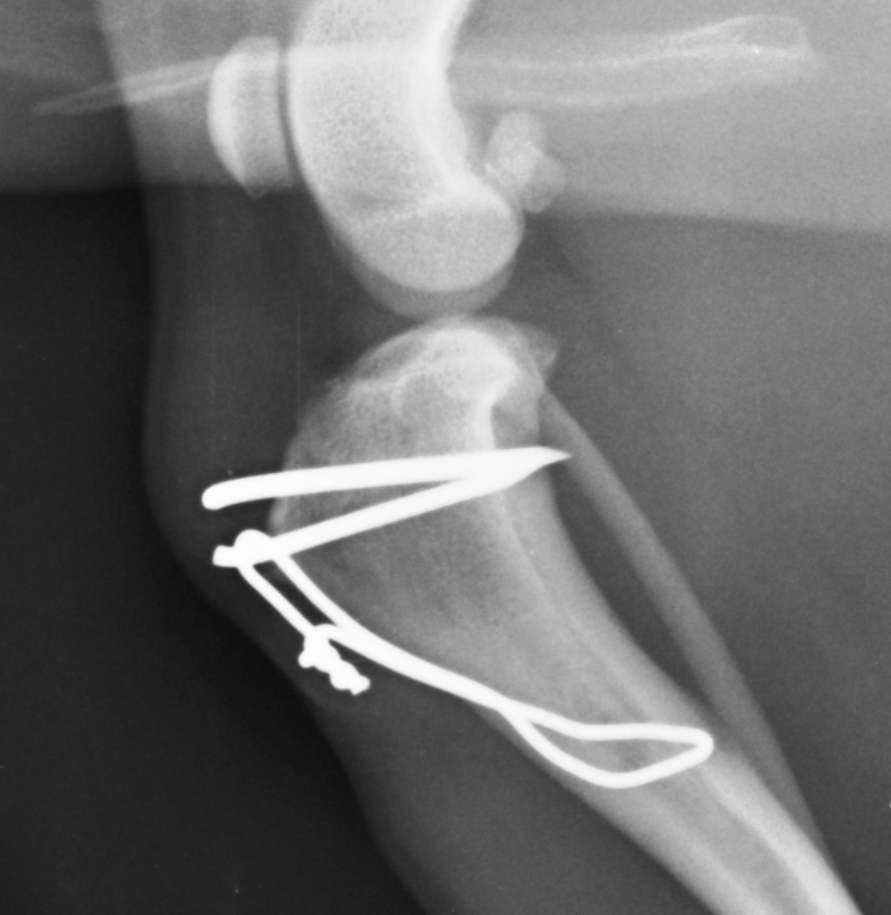
The aim of this surgery is to move a small piece of bone (the tibial tuberosity) at the top of the shin (tibia) that is attached to the patella and reposition it so that the patella is correctly aligned with the groove in the thigh (femur) bone. This procedure is called a tibial tuberosity transposition. The transposed piece of bone is re-attached with one or two small pins, with or without additional support with a figure-of-8 wire.
Exercise following quadriceps realignment surgery must be very restricted for the first few weeks until the cut bone and soft tissues heal. It must be on a lead or harness to prevent strenuous activity, such as chasing a cat or squirrel. At other times, confinement to a pen or a small room in the house is necessary. Jumping and climbing should be avoided. After a few weeks, exercise may be gradually increased in a controlled manner (still on a lead). Hydrotherapy may be recommended.
2. Trochlea (groove) deepening surgery
In dogs and cats with patellar luxation the groove (trochlea) at the end of the thigh bone (femur) is often shallow. In these cases it may be necessary to deepen the groove. This can be done by removing a block or wedge of bone and cartilage from the groove, deepening the base, and replacing the block or wedge. These techniques are called ‘recession’ techniques since they recess the surface of the groove and thus make the groove deeper, while at the same time preserving the surface (cartilage) of the groove.
3. Femoral osteotomy surgery
Femoral osteotomy surgery involves changing the shape of the deformed thigh bone (the femur) by cutting it just above the knee (stifle) and stabilising it in a new position with a plate and screws. This may be all that is needed to prevent the patella luxating, however, in some dogs it is also necessary to perform a tibial tuberosity transposition.
Exercise following femoral osteotomy surgery must be restricted until the cut bone has healed. Exercise must be on a lead or harness to prevent strenuous activity. Jumping and climbing should be avoided. X-rays (radiographs) are necessary between six and eight weeks following surgery to ensure bone healing is progressing without complication. Exercise may then be gradually increased in a controlled manner. Hydrotherapy may be recommended.
What is the outlook with patellar luxation surgery?
The outlook or prognosis with patellar luxation surgery is generally good. Although all dogs and cats develop osteoarthritis to some degree, this is often not a cause of pain or lameness. Stiffness, especially after rest, can be a feature in some cases. Potential complications include recurrence of the patellar luxation and loosening of implants. These are uncommon.