News
Archive for August, 2018
Perineal Rupture
by admin on August 31st, 2018
Category: News, Tags:
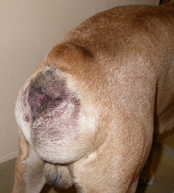
What is a perineal rupture?
A perineal rupture (sometimes called a perineal hernia) is a weakness or separation of the muscles of the pelvic diaphragm. The pelvic diaphragm is formed from a group of muscles that are situated around the rectum and form the caudal (back) wall of the abdominal cavity.
A perineal rupture is an abnormal hole in this diaphragm or sheet of muscles that allows fat or organs from the abdominal cavity to bulge into the area surrounding the anus (the perineum).
What are the clinical signs?
Clinical signs can include:
- Perineal swelling – this can fluctuate in size
- Perineal bruising
- Pain/discomfort associated with the swelling
- Pain or discomfort when passing of faeces
- Constipation
- Urine retention – an inability to empty the bladder properly
- Acute illness due to obstruction of urine flow from the bladder
- Generalised malaise
What animals are affected?
The condition predominantly affects middle aged to older male dogs, particularly entire (uncastrated) males. However, younger male dogs, female dogs and cats can also be affected.
What are the causes?
There is a strong association between perineal rupture and the male hormone levels in middle aged to older entire male dogs. Any disease process that affects the levels of male hormones can therefore predispose to perineal rupture.
In addition, any condition that results in straining or increased intra-abdominal pressure can result in failure of the pelvic diaphragm muscles.
Risk factors for perineal rupture include:
- Entire (uncastrated) male status
- Disease of the testicles (e.g. tumours)
- Disease of the prostate gland, which is found near the neck of the bladder in male animals (e.g. benign enlargement, abscess, cysts, tumours)
- Straining due to abdominal disease (e.g. colitis [inflammation of the large intestine])
- Pregnancy (due to increased abdominal pressure)
What does investigation involve?
In order to maximise the chance of successful treatment of the rupture, it is important to identify risk factors that are present in the patient and treat these at the same time as treating the rupture. Each investigation is likely to be tailored specifically to the patient, although typically this may include the following:
- Rectal examination (which may require sedation or general anaesthesia) – this will include assessment of the side opposite to the obvious rupture
- Blood tests
- Urine analysis
- Ultrasound scan of the abdomen and possibly of the hernia
- Prostate gland investigation/biopsy
- Examination of the testicles
- Chest X-rays in some cases
Cases with displacement of the bladder can present as emergencies and require urgent stabilisation prior to investigation and treatment.
What treatment options are available?
Most cases will require repair of the rupture and this is combined with castration in entire males to reduce the risk of failure. Those cases with other disease processes that have contributed to the development of the rupture will need to receive treatment for that underlying problem, prior to or at the same time as repair of the rupture.
Some ruptures are repaired using the muscles that are present within the pelvic diaphragm, although many require movement of additional muscle into the area in order to close the rupture. In some severe cases, mobilisation of a muscle from a hind leg or a synthetic mesh may be required to complete the closure.
Further support for the repair can be given by fixing the bladder and colon (large intestine) into the abdomen but this is not necessary in all cases.
What is the prognosis (outlook)?
Over 90% of uncomplicated cases receiving rupture repair and castration resolve following the surgery.
In experienced hands, complications following surgery are infrequent, and animals usually stay in the hospital for a few days to ensure they are comfortable and passing faeces easily.
Despite careful selection of the technique used and meticulous care, complications can occur in a minority of cases and these include:
- Recurrence of the rupture – stool softeners may be advised long term to reduce the straining during defaecation and therefore protect the repair
- Rupture of the other side
- Infection (the surgical site is in the perineal region, near to the anus which is a source of bacteria)
- Faecal incontinence – this is uncommon and usually temporary
Pet of the month – September – Mawgan
by admin on August 31st, 2018
Category: Pet of the Month, Tags:
Pet of the Month for September is a delightful Golden Retriever called Mawgan, seen here recovering from surgery in the lap of one of our nurses.
He came in to the clinic for our August Offer which was designated Check-a-Lump Month and subsequently underwent surgery to remove 2 lumps from his feet.
We are pleased to report that Mawgan is recovering very well!

Special Offer – September 2018
by admin on August 31st, 2018
Category: Special Offers, Tags:

Special Offer – August 2018
by admin on August 1st, 2018
Category: Special Offers, Tags:

Pet of the month – August – Max
by admin on August 1st, 2018
Category: Pet of the Month, Tags:
Max is recovering from a severe anaemia caused by a flea infestation. The severity was in fact so great that Max had to have an emergency blood transfusion, and his coat was completely shaved off.
Fleas are a blood sucking parasite and in sufficient numbers can deplete the number of red blood cells circulating in the body. The very prolonged warm spell we are having is causing flea numbers to rise exponentially. Each flea can lay up to 50 eggs daily and these can hatch within 14 days in hot weather. Although it is extremely unusual for a pet to be parasitised by fleas to this extent it is a warning to us all to maintain flea and all anti-parasite control this summer.

Pododermatitis
by admin on August 1st, 2018
Category: News, Tags:
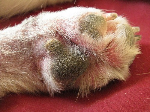
What is pododermatitis?
Pododermatitis is a term used to describe inflammation affecting the skin of the feet. It often causes dogs to have swollen, red and itchy feet, which can progress to painful sores if left untreated. In very severe cases, dogs can even become lame. This is a relatively common skin problem in dogs and can be present on its own or as part of a more widespread skin problem. There are many causes of pododermatitis and in some patients more than one cause is present at the same time. It is important to obtain an accurate diagnosis so that the right treatment can be selected.
What causes pododermatitis?
Parasites
The parasitic mite Demodex can infect the haired skin of the feet and result in pododermatitis. Demodex mites are present in very low numbers in the skin of all dogs, but in some patients, either due to a genetic susceptibility, or due to a process that lowers the immune system, these mites can populate the skin in very large numbers causing disease. Pododermatitis due to these mites tends to result in hair loss, swelling and bleeding sores in some cases. This mite is not infectious to other animals or people, but requires specific treatment to reduce mite numbers down to normal levels again. Very rarely, other parasites can also contribute to pododermatitis.
Foreign bodies
Foreign bodies like grass seeds are a very common cause of pododermatitis in dogs. Foreign bodies tend to penetrate the skin of the feet when dogs are out walking/running and then trigger inflammation when they become trapped within the feet. The body often attempts to ‘expel’ these structures resulting in painful and often discharging lumps between the toes. Affected patients often lick and chew at the affected sites. Foreign bodies are particularly likely when one lesion is present on one foot.
Allergies
Allergic diseases in dogs and cats are very common, and results in inflammation in the skin. This inflammation is very commonly seen affecting the feet, and results in redness, excessive licking and chewing at the affected sites. The most common triggers for allergic pododermatitis are food items and environmental substances such as dust mites and pollens, and skin disease usually starts in early life between the ages of 6 months and 3 years.
Deep infections
A very common feature of pododermatitis, particularly in dogs, is a deep infection of the feet. This is usually due to bacteria, but can be due to rare fungal organisms, and often results in multiple painful, swollen and discharging lumps. Affected animals usually lick and chew excessively and can become lame in severe cases. Bleeding lesions are relatively common with deep infections.
Conformation
A frustrating cause of pododermatitis is termed conformational pododermatitis. This usually occurs in heavy set dogs with excessively splayed feet. This results in weight bearing on hairy parts of the foot adjacent to the footpads and triggers inflammation of the hair follicles. Over time, this inflammation damages the hair follicles and results in chronic inflammation with the feet. Dogs with this condition tend to have large areas of pad extension, with painful and swollen lumps around the toes.
Hormonal diseases
Certain hormonal diseases can also be involved in the development of pododermatitis as the local skin immune system is reduced and the ability to fend off infections is compromised. The most commonly involved diseases include an underactive thyroid gland (hypothyroidism) or overactive adrenal glands (Cushing’s disease). However, pododermatitis is a relatively rare symptom of these diseases and dogs and cats usually show other more characteristic symptoms.
How is pododermatitis diagnosed?
Diagnosis of pododermatitis can often be achieved following a thorough evaluation of the history and clinical signs. Hair plucks and skin scrapings are performed to diagnose Demodex mite infestation and swab samples are often taken to establish if an infection is present. If lumps are discharging fluid, a sample of this fluid may also be sent to a laboratory to grow (culture) any infectious organisms. If the clinical picture is very suggestive of a foreign body, X-rays may be needed along with a surgical procedure to remove the offending item. Allergies are often only diagnosed once infections/parasites are treated and removed. If redness and inflammation remain, allergy testing may be required. Hormonal diseases are often suspected if other clinical signs are present but usually require blood testing to diagnose. Conformational pododermatitis is usually diagnosed by examining the feet and assessing the shape (conformation) of the footpads.
What are the treatments available?
Treatments for pododermatitis vary depending on the underlying cause. Parasitic infestations are usually treated with dips/rinses for the feet. Deep infections are often treated with long courses of antibiotics or antifungal medications in the rare cases due to fungal infection. Foreign bodies are best treated by identifying the foreign body and removing it in a minor surgical procedure. Hormonal diseases require treatment specific to the condition, but sometimes involve supplementing with hormone, as is the case in hypothyroidism. Allergic diseases are treated by identifying the triggers and removing them if possible.
Conformational pododermatitis is perhaps the most difficult to treat, as the defect is due to the conformation of the patient. Many of these cases can only be managed rather than cured and require modifications such as protective boots, good foot hygiene and avoidance of rough and uneven terrain. In some of the very worst cases, conformational pododermatitis can be corrected with surgery to fuse the toe webs together.
What is the prognosis?
As there are numerous causes of pododermatitis and more than one can be present at the same time, a good prognosis depends on identifying all the contributing factors and correcting them if possible. If this can be done, the vast majority of cases will have a good outcome. Cases of conformational pododermatitis are rarely cured, and require long term management.