News
Archive for November, 2018
Cataract Surgery
by admin on November 1st, 2018
Category: News, Tags:
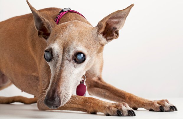
A cataract is an opacity or clouding in the lens in the eye. The opacity normally makes the lens look white. The lens in the eye is like the lens in a camera except that, rather than being at the front as it is in a camera, the lens in the eye is deep inside it, just behind the coloured part of the eye (the iris). The lens shows up as black in the central part of the eye (the pupil). The lens is normally crystal clear but it looks black because the darkness inside the eye shows through it. The lens is there to focus light on the sensitive tissue at the back of the eye (the retina).
Cataracts often form in both eyes and they frequently get worse. One eye is often more affected than the other, at least initially. It is not known why most cataracts develop. They are most common in older dogs and sometimes they occur due to other problems such as diabetes or disease in the back of the eye (the retina). Some cataracts are inherited.
What treatment is there for cataracts?
At the moment the only treatment for cataracts is surgery. Unfortunately not every cataract is suitable for surgical treatment, and it is necessary for the eye specialist to assess each patient and decide what therapy might be possible. Some cataracts are more complicated to treat than others, and the specialist will give guidance depending upon the circumstances in each case.
When is the best time to operate on a cataract?
The previous belief that it is best to let a cataract ‘ripen’ and the eye to become totally blind before removing the opaque lens (the cataract) has been proven wrong. Any cataract that is developing will cause potentially damaging inflammation in the eye due to release of lens proteins – a condition called ‘lens-induced uveitis’. Lens-induced uveitis can be subtle and easily missed – or, in some cases, it can be severe and associated with an obviously sore and inflamed eye.
Left untreated, even low levels of lens-induced uveitis are likely to result in complications, including adhesions (sticking) between the iris and the lens, retinal detachment (where the light-sensitive tissue at the back of the eye comes away from the back of the eye and stops working) and glaucoma (increased pressure in the eye which is potentially blinding and painful). Many eyes with long-standing cataracts that have not been operated on will eventually become irreparably blind and painful, and have to be removed as a result of the effects of lens-induced uveitis.
Early cataract surgery is therefore recommended in order to avoid the detrimental effects of lens-induced uveitis, and in general surgery is carried out once a cataract starts to interfere significantly with vision. This especially applies to young and diabetic patients, where progression of cataracts can often be rapid and result in significant complications if treatment is delayed. We will endeavour to see such patients at short notice and we will also often advise the referring veterinary surgeon to start treatment with topical anti-inflammatory drugs to manage lens-induced uveitis prior to us seeing the patient.
Cataracts in older dogs or patients with very slowly progressive cataracts may be monitored, but we will often recommend the prophylactic use of anti-inflammatory drugs in such cases.
The surgery
Your dog will usually be admitted on the morning of the surgery and no breakfast should be given. Water should not be withheld overnight. Diabetic patients need special management, and this should be discussed with the vet involved. After the patient is admitted, drops are given every 15 minutes prior to surgery to prepare the eye for the operation. These drops help to dilate the pupil and reduce the effects of inflammation which always happens in dogs having cataract surgery.
Before surgery all patients have an ultrasound scan to check for problems such as retinal detachment or rupture (bursting) of the lens. These changes are more common in advanced cases. The scan is performed under sedation. Some patients may be found to be unsuitable for surgery when the ultrasound scan is carried out.
Cataract surgery is performed under a full general anaesthetic and a muscle relaxant is given so that the eye comes into the correct position for the operation. This means that a ventilator needs to be used to inflate the chest during the procedure. We monitor your dog very carefully throughout the surgery using very modern sensitive equipment and the staff involved are specially trained in the procedure. This helps to reduce the risks of the anaesthetic to a very low level.
The operation is very delicate and involves the use of an operating microscope and tiny instruments. Two small cuts are made in the window of the eye (the cornea), near where the coloured part (the iris) joins the white part.
The lens is just behind the iris and lies in a delicate bag of tissue called the capsule. After the eye has been filled with a special gel called a viscoelastic, some of the lens capsule is taken out. The gel which is used helps to inflate the eye and protect the structures inside it from the effects of the surgery, and especially from the instruments and the ultrasound. The cataract (in other words, the lens) is then removed through the hole in the capsule using a technique called phacoemulsification – this is an ultrasound procedure using very sophisticated equipment which is exactly the same as that which would be used on a human eye. This type of surgery has been shown to give the best results in dogs’ and humans’ cataracts. There is currently no laser treatment for cataracts in dogs or humans.
In most patients it is possible to put in a special artificial lens where the old lens was. Plastic lenses make vision in the eye similar to the way it used to be before the cataract developed. The lenses used in our patients are made especially for dogs as they are bigger and more powerful than human lenses. They are permanent and buried deep inside the eye. The complication rate of lenses is very low indeed. For technical reasons it is not possible to implant an artificial lens in some eyes. Not having a lens implanted does not make the difference between being blind and having sight – it is similar to someone who wears glasses not putting them on.
At the end of the surgery the wounds in the eye are closed with tiny dissolving stitches. These are absorbed over the next few weeks, leaving only very small scars.
Some dogs can have both eyes operated on at the same time. The main reason for doing this is that it makes it more likely that the patient will have vision after the surgery – if something goes wrong with one eye, hopefully it will not also go wrong in the other one. However, a dog with one good eye will have overall vision which is almost as good as that in a dog with two good eyes, and so it is not essential to have both eyes operated on.
Most patients stay in overnight after their operation and are discharged the following day, provided that progress is satisfactory. Most dogs can see something on the day after surgery, but it frequently takes a few weeks for vision to settle down as the eye adjusts to the effect of surgery and the presence of a plastic lens implant. In addition, there is often some clouding inside the eye which takes time to clear.
Aftercare
The aftercare following cataract surgery is intensive. All patients develop inflammation inside their eyes after surgery. This happens more in dogs than in humans. Usually there are several types of drops used. The most frequently applied drops are used six times daily initially. The number of applications gradually decreases over the next two months or so. There are also tablets to be given for a few weeks after the surgery.
Your dog will need to be kept as quiet as possible for a few weeks after the surgery, although this can obviously be difficult with many of our patients! You can only do your best in this regard. Pulling on a lead should be avoided for several weeks after surgery, as this puts up the pressure inside the eye and can encourage bleeding. Avoiding pulling around the neck is best achieved by using a harness, and it is a good idea to obtain one before the operation – it can be fitted at the time that your dog goes home. A plastic Elizabethan collar also has to be worn for about a week after the operation.
There will need to be at least four or five re-examinations after surgery. These are mostly within the first two to three months after the operation. Some patients, especially those with complicated cataracts, may need longer term treatment and more check-ups than average.
Risks and complications
The success rate of cataract surgery in dogs is about 90 to 95% initially. This means that 5 to 10% of patients cannot see in the operated eye after surgery. There are various reasons why not all patients have a successful outcome or may have a less straightforward recovery than normal. These include:
Inflammation
Every patient gets inflammation after surgery, no matter how smoothly the surgery goes. This is usually well controlled by the medications which are given. The occasional dog gets more inflammation than average, and this can lead to changes in the eye. These may not be of any great significance, but sometimes they can cause reduced vision.
Occasionally an injection into the eye is needed to dissolve inflammatory clot material. Inflammation is the main problem in dogs after surgery, and is the major reason why frequent medications and regular post-operative check-ups are required.
Infection
This can be very serious, but is extremely rare. Antibiotics in the form of tablets and ointment are used before and after the surgery to help to prevent this.
Wound breakdown
This means that the wound gives way. Again this is an uncommon complication, but if it occurs another general anaesthetic will be required to re-stitch the wound.
Bleeding
A very small amount of bleeding at the time of surgery is not unusual and this is not a major problem. Very occasionally a larger haemorrhage can develop and this can affect vision.
Increased pressure
The pressure in the eye can occasionally go up in the first few days after surgery, but eye drops will usually settle this down very quickly. Rarely a more severe increase in pressure may develop (glaucoma). If this problem develops it will involve additional medication and possibly surgery. It can lead to blindness and even loss of the eye in severe cases which don’t respond to treatment.
Ulcers
Occasionally the surface layer of the window at the front of the eye (the cornea) can partly come away after surgery. This is usually a very minor problem which normally resolves within about a week.
Corneal oedema (water-logging)
The window at the front of the eye (the cornea) can very occasionally go blue after surgery due to disturbance of its inner layer. Careful surgery and the use of viscoelastic gel (as previously mentioned) help to reduce the chances of oedema developing.
Retinal detachment
This is an uncommon complication, but if the sensitive tissue at the back of the eye detaches it can lead to loss of sight. A routine ultrasound scan before the surgery helps to identify at-risk patients.
Poor vision
Some dogs have problems inside their eyes (for example with their retina) which cannot always be detected before the surgery, and this may then mean that the surgery is not successful, or that the vision given by the surgery is not as good as it once was. Some suspect cases may have an electrical test (an electroretinogram) performed on the eyes to look for retinal problems before surgery, but this may well require sedation or even general anaesthesia and is not necessary or recommended for every case.
‘After-cataract’
A small percentage of dogs that see well immediately after their surgery may not continue to do so for the rest of their lives. This later deterioration may happen for many reasons (such as some of the complications mentioned). However, one such problem is known as after-cataract, in which a white membrane can grow across the pupil inside the eye. In most patients the amount of after-cataract which forms is not significant, but it can very occasionally affect vision in the long-term. Having a plastic lens implant has been shown to help to prevent the membrane growing across the pupil.
If after-cataract becomes very severe it can be removed surgically, although this is very rarely necessary.
Conclusion
The success rate of cataract surgery in dogs is high, and the great majority of patients do very well after their operation. It is undoubtedly a major undertaking, but the procedure is one which is commonly performed.
Pet of the month – November – Kim
by admin on November 1st, 2018
Category: Pet of the Month, Tags:
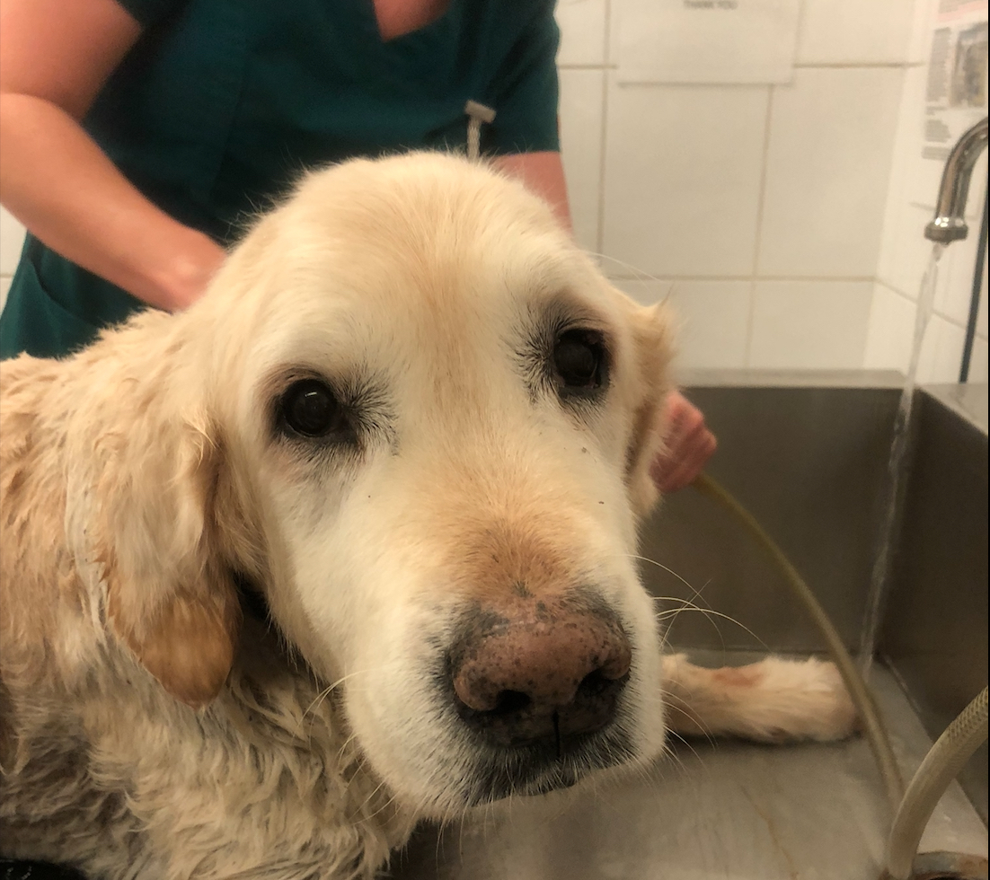
Pet of the month for November is Kim, a 14 year old Golden Retriever, seen here having her weekly bath. Kim has suffered from an underactive thyroid for some years and despite longterm supplementation with thyroid hormone still suffers from a flaky skin, which is a common symptom. Other than that the condition is being well managed.
What is hypothyroidism?
Hypothyroidism develops when the thyroid gland function falls below normal. In dogs and cats the two thyroid gland lobes lie on either side of the windpipe and produce thyroid hormone. This hormone controls the speed at which the body’s metabolism works. In patients with hypothyroidism, not enough of the hormone is produced. As a result the metabolism ‘slows down’, and, with time, this can affect almost all the organs of the body.
Hypothyroidism is common in adult dogs, but is very rare in cats.
How do animals get hypothyroidism?
In the majority of cases hypothyroidism is diagnosed in adult dogs that have previously been healthy. In most dogs the disease either develops due to the affected animal’s own immune system attacking the thyroid gland (‘lymphocytic thyroiditis’) or due to degeneration of the gland tissue. It is possible that both forms of the condition may in fact represent the same disease process. Over time, more and more thyroid tissue is destroyed until the gland cannot function any longer.
It is still not known why dogs develop this disease. Certain breeds of dogs are more susceptible to hypothyroidism – these include Retriever breeds, Doberman Pinscher, Setters, Terrier breeds and Spaniels (especially Cocker Spaniels). Hypothyroidism is rare in toy and miniature breeds. Several factors have been proposed that could trigger the immune reaction, but so far nothing has been proven.
Hypothyroidism is extremely rare in cats, but can very occasionally be seen after zealous treatment of an overactive thyroid gland.
In rare cases puppies and kittens are born without a functioning thyroid gland.
What are the clinical signs of hypothyroidism?
As the disease affects the whole metabolism, clinical signs are often vague and non-specific and can affect almost every aspect of the dog’s body. The disease can also mimic many other problems.
Many dogs are presented with weight gain and lethargy or lack of interest in activities they previously enjoyed. However, the majority of dogs presented because of weight gain are simply overweight because they eat too much (See Nutrition advice for dogs information sheet).
Often a ‘tragic expression’ of the face is noticed by owners of dogs with hypothyroidism, and this is due to thickening of the skin and loss of tone of facial muscles. A dull coat, greasy or darkened skin and hair loss, especially on the sides of the body and the tail, are also frequently noticed. Constipation can develop, although diarrhoea is also a possible problem because the immune system is less effective. This reduction in immunity can also lead to other infections such as chronic skin or ear problems. Less commonly, more severe clinical signs can be seen, such as those related to nerve function problems.
Hypothyroidism is not usually fatal, but it can compromise an affected dog’s quality of life.
Puppies and kittens born without a functioning thyroid gland do not grow properly and their mental development may be affected. Many of these patients do not survive, and those which do often suffer from chronic arthritis because of abnormal joint development.
Adult cats with hypothyroidism show similar signs to dogs, but such cases have only very rarely been reported.
How is the disease diagnosed?
Hypothyroidism is diagnosed by blood testing – the amount of thyroid hormone in the blood as well as the amount of TSH (a hormone that controls the function of the thyroid gland) is measured. However, further blood values are also looked at in order to test for other diseases, because the clinical signs of thyroid disease are not usually very specific. Depending on the individual case it may also be necessary to analyse a urine sample or even to perform further tests such as radiography (X-rays) or ultrasound examination.
If other diseases can be ruled out and the blood values show low thyroid hormone, then a diagnosis of hypothyroidism can be straightforward. However, not all blood tests results lead to a clear-cut diagnosis as thyroid hormone measurement can be influenced by a variety of other factors. In such cases it may be necessary to perform further blood tests, or in some cases to re-test after several weeks or months to confirm the disease. In some cases the diagnosis of hypothyroidism can be quite problematic.
Can hypothyroidism be treated?
Once hypothyroidism has been diagnosed, it can be readily treated with tablets containing thyroid hormone. The condition can be controlled rather than cured, and life-long medication has to be given to provide the body with thyroid hormone. As the required dose can change over time, repeat blood tests are necessary at intervals to check the level of hormone in the blood.
The clinical signs of hypothyroidism will improve with treatment but it can take several months for all the signs to resolve.
My pet has hypothyroidism, what is the outlook?
Once a pet is stabilised on medication, it will not be noticeable to anyone – including the animal – that he or she actually has a chronic disease. Pets with hypothyroidism that are on therapy have an excellent prognosis and can expect to live a normal life, as long as they receive medication and are properly managed with the help of your veterinary surgeon.
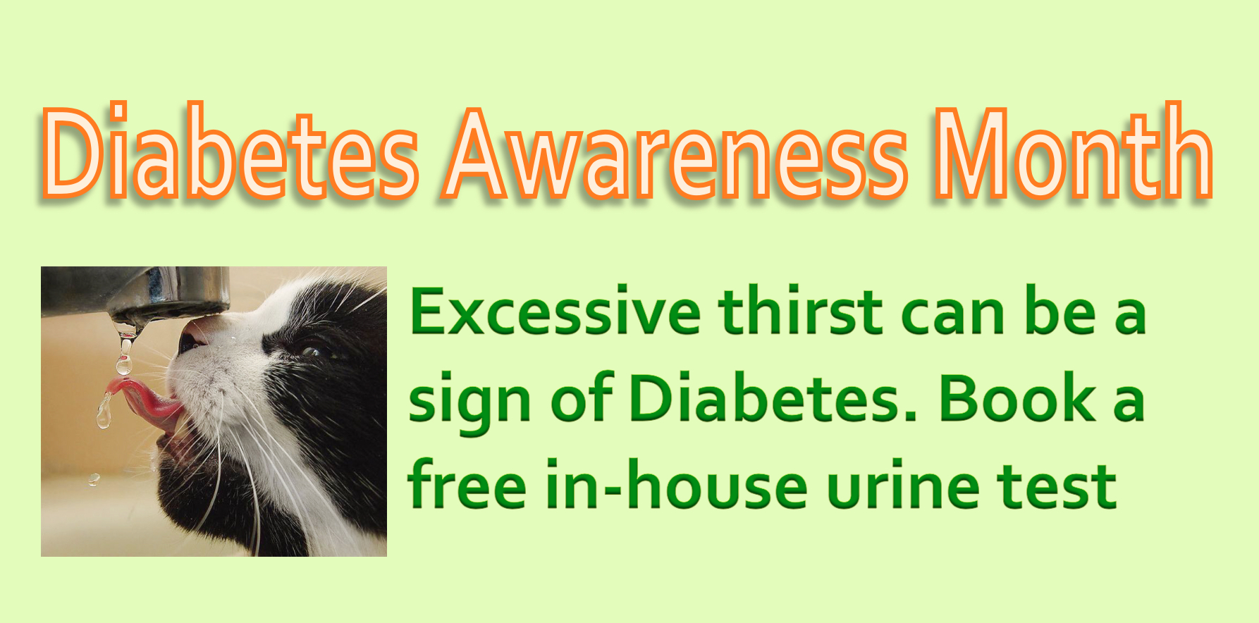
Special offer – November 2018
by admin on November 1st, 2018
Category: Special Offers, Tags: