News
Archive for August, 2017
Special Offer – September – Dental Offer
by admin on August 31st, 2017
Category: Special Offers, Tags:

Glaucoma
by admin on August 31st, 2017
Category: News, Tags:
What is glaucoma?
Glaucoma is an increased pressure inside the eye, caused by an obstruction to the drainage of the fluid from within the eye. In order to keep the eye inflated, fluid is normally produced and cleared away from the eye all the time. The fluid inside the eye is not related to the tear fluid (which is on the surface of the eye).
The blockage of fluid drainage within the eye can be due to an inborn defect (primary glaucoma) or due to another eye disease that interferes with drainage of the fluid inside the eye (secondary glaucoma). Common causes of secondary glaucoma are inflammation inside the eye or a shift of the position of the lens within the eye.
Is my dog at risk?
Many cases of primary glaucoma are inherited and due to an abnormally formed drainage passage within the eye. Affected breeds include the Basset Hound, Welsh Springer Spaniel, Cocker Spaniel, Siberian Husky, Great Dane, the Flat Coated Retriever and many others. Often one eye is affected initially but there is a high risk that the other eye will follow at some point in the future.
A test (gonioscopy) is available to determine the predisposition of your dog to develop glaucoma. In this test, a special contact lens is applied to the eye to allow assessment of the structure concerned with drainage of fluid from the inside of the eye.
What are the signs of glaucoma?
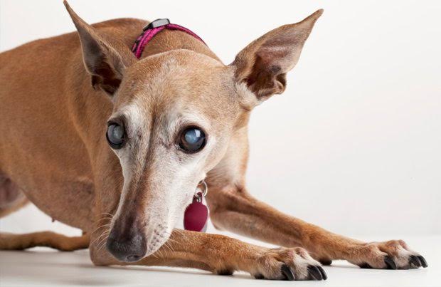
In most cases, the disease develops very rapidly. The patient is often depressed and reluctant to exercise. The eye becomes blind and appears red, painful and sore with a bluish tinge over the cornea. Less commonly, the pressure increase is slow and the clinical signs are not as pronounced. However, a gradual reduction in vision is often noticed.
How is glaucoma diagnosed?
A special instrument called a tonometer is used to measure the pressure inside the eye. Local anesthetic is applied to the eye for this test which is usually very well tolerated.
Is treatment possible?
The aim of treatment in glaucoma is to preserve vision and to relieve the pain caused by the pressure increase. In order to reduce the pressure within the eye, drugs are given to reduce fluid production within the eye and to improve removal of fluid from the eye. In some cases of secondary glaucoma (see previously mentioned), treatment of the underlying cause, such as anti-inflammatory medication or removal of a dislocated lens, can lead to the pressure decreasing.
Patients with primary glaucoma are more difficult to treat and it is important to realise that no cure for the disease exists. In some patients, pressure control can be achieved with medical treatment only. However, with time, most patients become less responsive to the treatment and surgical alternatives may have to be considered. These include laser therapy or the surgical placement of a drainage implant into the affected eye.
Regular check ups will also be necessary to re-assess the second eye and check its pressure.
Is preventative treatment available for the second eye?
No preventative treatment is available that can totally stop glaucoma developing in the second eye. However, there is evidence that with the help of medication, the onset of the condition in the second eye may be delayed.
What happens if the pressure cannot be controlled?
Eyes that have lost vision but continue to have an increased pressure are a cause of chronic pain for the patient. Removal of the eye must be considered in such cases to ensure the welfare and comfort of the patient. Occasionally, both eyes may, unfortunately, be lost. Should this be the case, most dogs will adapt very well to being blind and continue to lead a good quality life.
Syringomyelia
by admin on August 1st, 2017
Category: News, Tags:
Syringomyelia is a relatively common condition, especially in breeds like the Cavalier King Charles Spaniel and the Griffon Bruxellois, in which it is suspected to be an inherited disorder. Other names that have been used to describe this condition include syringohydromyelia, Arnold-Chiari or Chiari-like malformation, and caudal occipital malformation.
What is syringomyelia and what causes it?
Syringomyelia is a neurological condition where fluid filled cavities develop within the spinal cord (the bundle of nerves that run inside the spine). The most common reason for the fluid build-up is that there is an abnormality where the skull joins onto the vertebrae (the bones of the spine) in the neck, causing fluid in the brain (called cerebrospinal fluid or CSF) to be forced down the centre of the spinal cord, where it causes the tissues to become distended and cavities to form.
What are the most common signs of syringomyelia?
Clinical signs or symptoms can vary widely between dogs and there is no relation between the size of the syringomyelia (cavity in the spinal cord) and the severity of the signs – in other words a dog with severe fluid build-up can have relatively mild symptoms, and vice versa. The most common symptom that develops is intermittent neck pain, although back pain is also possible. Affected dogs may yelp and are often reluctant to jump and climb. They may feel sensations like ‘pins and needles’ (referred to as hyperaesthesia). Another common sign is scratching of the neck and shoulder region called ‘phantom scratching’, as there is generally no contact of the foot with the skin of the neck. Occasionally dogs become weak or wobbly if there is significant damage to nerves within the spinal cord. Cavalier King Charles Spaniels will typically show clinical signs between 6 months and 3 years of age. Not all dogs with syringomyelia will show signs of pain or other clinical symptoms, so the presence of syringomyelia can be an incidental finding on an MRI scan or specialised X-rays, when neurological investigations are being performed.
Other neurological conditions, such as slipped discs (cervical and thoracolumbar disc disease), can mimic the signs of syringomyelia and it is important for us to rule them out before concluding that your pet is suffering from syringomyelia.
How can syringomyelia be diagnosed?
The best method of diagnosing syringomyelia is an MRI scan of the brain and spine. It is necessary to perform this investigation under a general anaesthetic. The scan and anaesthetic are safe procedures. In the future it is possible there will be a genetic test to identify dogs with syringomyelia.
How can syringomyelia be treated?
Medical therapy is usually the treatment of choice in dogs suffering from syringomyelia. Several types of medication are used to manage episodes of pain, including a drug called gabapentin. This drug is safe, with few side effects apart from possible sleepiness. Other medications that may be used include anti-inflammatory drugs, corticosteroids and drugs that reduce the production of fluid in the brain and spinal cord.
Occasionally medical management is unsuccessful and surgery needs to be considered. The aim of surgery is to improve the shape of the back of the skull and reduce the flow of fluid down the centre of the spinal cord. Many dogs will improve following surgery, although some patients will have persistent signs despite surgery, whereas others may show improvement initially but then develop recurrence of their symptoms.

Pet of the Month – August 2017 – Harry
by admin on August 1st, 2017
Category: Pet of the Month, Tags:
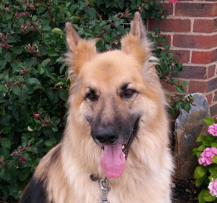
Pet of the Month this August is Harry, a handsome 7 year old German Shepherd Dog. We are delighted to report that he has recovered extremely well following recent surgery for Gastric Dilatation.
What is gastric-dilatation and volvulus (GDV)? Is my dog at risk?
Gastric dilatation and volvulus, or GDV as it is commonly abbreviated, is a relatively common clinical syndrome seen in large / giant breeds of dog. Dilatation refers to bloating of the stomach with gas, and volvulus refers to twisting of the stomach about its axis. The cause of this syndrome is not completely understood. In fact it is quite controversial which occurs first; the bloat or the volvulus (twisting). Indeed both components do not have to occur together and some patients will develop relatively simple bloat alone. GDV is a potentially life threatening condition and emergency veterinary attention should be sought immediately if it is suspected.
Why is GDV potentially life-threatening to dogs?
There are a number of serious and potentially fatal consequences that occur as a result of GDV. Initially the severe distension of the stomach stretches the blood vessels over its surface reducing the blood supply to the stomach walls. This is made worse by the twisting of the stomach which also twists the blood vessels, effectively shutting off blood supply to the stomach. A lack of blood flow means there is a lack of oxygen and nutrients delivered to the stomach and waste products are not removed. As with any organ this will result in parts of it dying. This process happens very quickly and in severe cases could result in part of the stomach wall rupturing and releasing its contents into the abdomen.
The large distended stomach occupies much more space inside the abdominal cavity and compresses surrounding structures. Severe distension puts pressure on the diaphragm and interferes with the patient’s ability to breath. It also applies pressure to a large blood vessel in the abdomen (the vena cava) that normally returns blood from the back half of the body to the heart. Pressure on this vessel obstructs flow therefore reducing the amount of blood returned. If blood can’t be returned to the heart, then it in turn can’t pump it out to the rest of the body. If there is insufficient blood being pumped, the blood pressure falls dramatically making the patient weak and potentially leading to collapse.
To add insult to injury, other organs in the body such as the lungs, kidneys, liver and intestines do not receive a blood supply and begin to fail. The lack of a functional circulation also means that toxic products build up in these organs that further compromise the patient. These changes can happen in a matter of hours, emphasizing the importance of early veterinary attention.
What are the symptoms of GDV in dogs?
The symptoms generally include obvious distension or enlargement of the abdomen with unproductive vomiting or retching. The patient may drool excessively and appear restless or agitated. As the condition progresses the patient may become increasingly weak or even develop shock and collapse.
What is the treatment for GDV?
The age and breed of the patient coupled with the clinical signs of a severely bloated abdomen will make your vet highly suspicious of this condition. They will immediately place one or more an intra-venous catheters to allow administration of fluids to support the circulation and dilute toxins in the blood. They may also analyse the patient’s blood to assess the severity of organ damage.
The next stage involves attempts to decompress the stomach. This is usually accomplished by passage of a specially designed tube through the mouth down into the stomach. There is a gag that can be used to assist in this process but many patients will require sedation or anaesthesia to complete the task. It can be very challenging or sometimes impossible to perform stomach tubing. This is particularly the case when the stomach is twisted 360 degrees or more. In this instance a cannula (tube) may have to be inserted through the body wall and into the stomach to allow deflation. Deflation is clearly an imperative step because it will relieve pressure on the diaphragm and help restore blood flow back to the heart through the vena cava.
Radiographs of the abdomen are often required to help distinguish between simple dilatation and dilatation with volvulus. In the latter case surgery will be required as soon as the patient is stabilised. The aim of the surgery is to de-rotate the stomach and assess it for areas of devitalisation. If there are areas of the stomach that have undergone necrosis (died), these need to be removed surgically.
It is vital that the stomach is attached to the inside of the body wall. This is called a gastropexy and it will prevent volvulus in the future. This is essential as up to three quarters of the patients that do not have this performed will have another episode in the future. This also applies to those patients suffering with bloat alone as they have the same risk.
What are the risk factors for GDV in dogs?
There are several factors that have been clearly demonstrated to increase an individual’s risk of developing this condition. These include:
- Being a purebred large / giant breed
- Having a deep and narrow chest conformation
- Having a history of previous bloat
- Having a history of bloat or GDV in a first degree relative (parent or sibling)
- Increasing age
- Having an aggressive or fearful temperament
- Eating fewer meals per day
- Eating rapidly
- Being fed a food with small particle size
- Exercising or stress after a meal
What breeds are predisposed to this condition?
The breeds most commonly affected are large purebred dogs that have a narrow deep chest confirmation. Those at most risk include:
- Great Danes
- Gordon setters
- Irish setters
- Weimaraners
- St. Bernard’s
- Standard poodles
- Bassett hounds
Although these are the breeds we typically see GDV in, it is worthy to remember that it can happen in any patient.
What is the prognosis?
With improved understanding of the secondary consequences of GDV and excellent anaesthesia, surgical and post-operative care now available for veterinary patients a good prognosis can be achieved for this condition. Survival rates of 73-90% would be typical. There will always be a range quoted for survival because individual patient’s circumstances in terms of severity, age, general health and treatment received will have an impact on the outcome. One important factor that has been shown to decrease the survival is the presence of clinical signs for greater than six hours. This emphasizes the importance of prompt veterinary attention in all cases.
If you are in any doubt that your dog is suffering from bloat or GDV, please call your vet immediately.
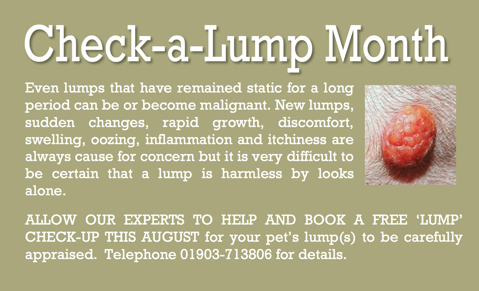
Special Offer – August – Check a lump month
by admin on August 1st, 2017
Category: Special Offers, Tags: