
News
Archive for the ‘News’ Category
Episodic Weakness and Collapse
by admin on July 1st, 2016
Category: News, Tags: Tags: collapse, dog, weakness

What is Episodic weakness and collapse?
Essentially, ‘episodic weakness and collapse’ refers to involuntary falling over! Dogs are more commonly presented to veterinary surgeons with this problem than cats. Syncope (pronounced sin-coh-pea) is the technical term for fainting when the patient temporarily loses consciousness. Collapse, such as fainting, may be completely benign and require no treatment. However, in some circumstances it is due to a life-threatening situation that requires a specific treatment.
Remember these conditions are both Involuntary (the patient has no control over them) and intermittent or ‘episodic’ and the patient may be completely normal between bouts. There are no seizures or fits as we see in Epilepsy. Also, before starting our investigations, we need to be as sure as possible that involuntary collapse is truly occurring. If a dog is choosing to lie down, for instance because of tiredness or heat exhaustion, or because of feeling faint after pulling hard on the lead this is not collapse.
What causes episodes of weakness, collapse or fainting?
The causes are many and varied and can involve:
- The airways
- The heart and blood vessels
- The nervous system – brain, spinal cord, peripheral nerves
- The muscles, bones and joints
- A malfunction of one or more chemical processes occurring in one of a number of body organs (these are usually referred to as ‘Metabolic’ causes).
To further complicate matters there are plenty of potential causes of collapse within each of these broad categories. So, what might seem to be a relatively ‘simple’ clinical presentation can turn into the proverbial search for a needle in a haystack when it comes to finding out the cause of the problem.
Investigations can therefore be costly, frustrating and extremely time consuming for all concerned. For example, it is quite common for blood samples to be sent to more than one specialist laboratory and some of these may be outside the UK. Finally getting all the results together can take several days or even weeks. It can be a trial of patience waiting for such tests but it is better to wait for accurate results than to choose a more rapid but less helpful alternative.
How can pet owners and carers help in getting to a diagnosis?
Getting to a diagnosis is clearly very important so we know how to treat effectively. You can help your vet a lot in this regard! It is often very helpful if video footage of the events occurring can be provided as sometimes this may show important information which to the untrained eye may not be obvious. Your vet may well not see the event you are concerned about as by their very nature these are intermittent events.
It is very helpful to know the following:
- What a pet is doing just before he or she collapses
- What he or she does during the collapse
- What he or she does after the collapse
Your vet will then want to perform a thorough clinical examination. Ideally, the information you provide and the results of your vet’s examination will give diagnostic ‘clues’ as to which direction in which to look first. If these findings make one particular cause of collapse more likely than others then this gives a far better chance of determining the cause. A problem arises where signs are vague and do not allow us to ‘home in’ on a particular group of possibilities. In these circumstances unfortunately investigation needs to be very broad-based and we would start by evaluating for the most common causes of collapse.
How is collapse investigated?
There is no single test that will evaluate the patient for all causes of collapse. Tests are picked on an individual basis according to how valuable the clinician expects the test to be in a particular case. This is why the intial information and history is so important because it will help the clinician to be more specific with the tests performed and therefore more likely to discover the cause.
Each case is different but investigations will often involve
1. Blood tests to look for metabolic causes
2. Assessment of the heart by an electrocardiogram (ECG), an echocardiogram (heart ultrasound) and chest x-rays. In some patients where an intermittent electrical abnormality is suspected, the patient will be fitted with a device to perform an ECG for 24 hours in an effort to ‘catch’ the irregular heart rhythm. This special ECG device is called a Holter monitor and it is connected to sticky pads which adhere gently to the skin to allow the rhythm of the heart to be recorded for extended periods of time and even at home.
If these tests are normal then further testing depending on the remaining clinical possibilities is indicated. Sometimes further tests may be advised straight away or in some instances it may be worth performing these after a delay both to make sure that the problem is persisting and also to allow some time for further ‘clues’ pointing to a particular diagnosis to develop.
Is a specific diagnosis always found?
In some instances, despite lengthy and thorough investigation, no cause of the collapse is found. The same is also true in human medicine. Sometimes this may be because the cause is either benign or occurring so rarely that the cause is not caught ‘in the act’. Other causes of collapse may remain undiagnosed because some potential causes are simply not possible to evaluate in dogs and cats, particularly tests that in humans would require a period of voluntary bed rest – clearly this is not possible to achieve in a dog or cat.
How is Episodic weakness and collapse treated?
This completely depends on identifying the cause. Appropriate treatment is then instigated.
Management of Atopic Dermatitis in Dogs and Cats
by admin on June 1st, 2016
Category: News, Tags: Tags: atopic dermatits, cats, dogs, pets
Last month we explained what atopic dermatitis is and how it is diagnosed. In this article we consider how atopic dermatitis can be managed in affected dogs and cats.
Can I cure atopic dermatitis?
In one word “No”, but you can manage the condition successfully. Atopic dermatitis in dogs and cats can be compared to asthma in people. Asthma can’t be cured but it can be managed; and just like asthma the management of atopic dermatitis is life-long. It is therefore important to put in place measures that are going to have the least side-effects for your pet in the long term that will provide him/her a good quality of life and that are the most cost effective and affordable for you.
How can I manage the condition?
You need to take a multi-step approach to managing atopic dermatitis. These steps include:
- Treatment and prevention of infections
- Medications to stop the itch
- Treatment that modulates the immune system
- Nutritional supplements
- Topical treatments
- Unfortunately some treatments work some of the time but not all the time and so you may need to switch treatments periodically. Some treatments may result in undesirable side effects and so may need modification.
Management options:
1) Treatment and prevention of infections:
Any bacterial (mostly staphylococcal) and/or yeast (Malassezia) infections should be treated at the outset. Antibiotics for bacterial infections need to be administered for 7 – 14 days beyond clinical cure and the duration of treatment is generally determined by the depth of the pyoderma.
For yeast infections a shampoo containing either 2% chlorhexidine/2% miconazole or one containing 3% chlorhexidine have shown good efficacy and will suffice; however, if the infection is severe your vet may opt to prescribe antifungal tablets. Because there is a tendency for infections to recur it is best to bath your pet once or twice a week on a regular basis, which will keep the microbial load on the skin low and thus help reduce the frequency of infections.
2) Stopping the itch:
Glucocorticoids (steroids) will stop the itch in most cases, but need to be used with caution, especially if used for a long time. In the short term they can increase water intake, increase urination and appetite. Some owners and pets find these side effects distressing. In the long term they can affect almost any organ in the body, therefore, even when well tolerated, they should be used with caution and your pet should be monitored regularly. Topical steroid containing sprays and ointments may be useful for targeted areas but with long term use thinning of the skin, infections and systemic side effects can occur.
Cyclosporin is used to damp-down (modulate) an over-reacting immune system in an atopic pet. One starts with a high dose and then tapers it down to alternate day, or better still twice a week, treatment. The most common side effects with ciclosporin are vomiting and diarrhoea. In some dogs, gingival hyperplasia and papillomas may also occur.
Oclacitinib is another immunomodulating drug which specifically targets the pathway that results in an itch. It is effective and can stop itching rapidly. This is the newest drug on the market and the reported side effects are vomiting and diarrhoea in a small number of cases. It can be used both short term and long term; however, given that this drug has now been on the open market for only a few months (at the time of writing this) it should be used with caution as side effects with prolonged use are not known.
Antihistamines; the response to this group is variable and they are often used in combination with steroids in order to reduce the steroid dose.
3) Immune Modulation:
Allergen specific immunotherapy is the most specific treatment for Atopic Dermatitis. It involves either an injection, or an oral dose of the allergens your pet is allergic to, to modulate the immune system. It has responses ranging from roughly 33% where there is complete cessation of itch, to 33% improving but requiring additional treatments, to 33% where there is no response. If your pet responds well to immunotherapy, it is likely to have the least adverse side-effects and to be the most cost effective treatment in the long term.
4) Nutritional support:
Omega -3 and Omega -6 essential fatty acids help the skin function by altering the lipid barrier and by reducing inflammation. On their own they are unlikely to benefit your pet but can be helpful when combined with other treatments. Diets that are high in essential fatty acids (EFAs) also help.
5) Topical treatments:
Moisturisers containing ceramides help maintain the skin barrier, which in turn reduces the penetration of allergens and therefore your pet’s reaction to them. Bathing with an oatmeal based shampoo can also help relieve itching and re-hydrate the skin.
Atopic dermatitis is a lifelong condition that requires life-long management; and often more than one type of treatment will be required. So, when your vet chooses a treatment, it should be efficacious, suitable for your pet with the least side effects, easy for you to administer and affordable. Most of all it/they should improve your pet’s (and your) quality of life.
Atopic Dermatitis
by admin on May 2nd, 2016
Category: News, Tags: Tags: atopic dermatits, dog, skin, sore
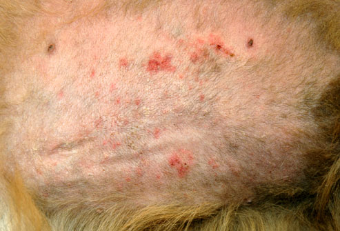
What is atopic dermatitis?
Many of you will have been told by your vet that your pet has atopic dermatitis, but what is it? Atopic dermatitis is the visible sign of a hypersensitivity (allergic) reaction; specifically a reaction to something (called an allergen) in your pet’s environment, either indoors or outdoors. It may be a reaction to pollens, or moulds, or to mites, such as house-dust mites.To complicate the issue further, some pets can have an allergic reaction to some foods, which is ‘food induced atopic dermatitis’.
Hypersensitivity is when the immune system goes into an overdrive. Normally when a pet is exposed to an allergen, its immune system produces antibodies to the allergen and once it has performed its protective role it will automatically revert to normal; however, if your pet is predisposed to atopic dermatitis its immune system continues to produce antibodies, which may then result in a hypersensitivity.
What are the main signs of atopic dermatitis in dogs and cats?
The hallmark of atopic dermatitis is an itch, usually without any visible signs other than a reddening of the skin. Often when I ask an owner where their pet animal is itching they say “Oh, Fido does not scratch”, but dogs exhibit itch by licking, rubbing, or chewing at the itch as well as by scratching at it.
The itching usually starts between the ages of 6 months and 3 years. Initially it may be seasonal – that is it occurs only at certain times of the year – but over the years that can change. Spring and summer time itching is usually associated with pollen allergies (e.g. tree, grass and weed pollens) whereas winter or all year round itching is associated with indoor allergens such as house dust and storage mites or mould allergens. Food associated atopic dermatitis is likely to be non-seasonal, unless your pet is only being fed the food intermittently (e.g. on holiday). The itchy areas of the skin are usually the face, ears, feet and underside.
Often the condition is complicated with secondary infections (bacterial and/or yeast) and sometimes, just to complicate the situation further; some patients have been known to develop an allergy to the infection as well. Infections increase the level of itching and so just treating these will often decrease or stop the itch entirely. Once infections occur a rash may appear on the belly, neck and feet and your pet will lick, chew or scratch these areas. Infected skin is malodorous (has a bad smell) and, as the infection progresses the skin becomes thickened, blackened and crusty. If your pet then licks or scratches these areas aggressively, the skin can become broken or ulcerated, and also bleed.
Since similar lesions can also be seen with parasitic infestations (e.g. mange) these must be ruled out, or treated, at the same time as the allergy.
Recurrent ear infections are also associated with food or environment associated atopic disease. Usually the ear flap and the ear canals appear red in the early stage, which is then often followed by head shaking, discharge and swelling.
What is my pet allergic to?
To find out what your pet is allergic to, your vet can perform an allergy test, which will either involve sending a blood sample to a laboratory, or performing an intradermal skin test. Intradermal skin tests are when very small amounts of allergens are injected under the skin to identify which ones that particular animal is allergic to by provoking a small, controlled allergic response. Both forms of testing have their advantages and disadvantages. The results of the test should be interpreted bearing in mind the seasonality of the condition and your pet’s environment.
Allergy testing identifies the allergens to which your pet might be allergic to, but the main reasons for testing should be to find out what your pet is allergic to, so as to be able to improve quality of his/her life. Your vet can use the information for therapeutic purposes and to look at means of reducing the allergen load in the environment. The lower the allergen load, the lesser the itch and the easier it will be to keep your pet comfortable. One can compare atopic dermatitis in dogs to hay fever in people. If you have hay fever and you stand in a field full of flowers whose pollen you are allergic to, you will suffer severely, as opposed to if you stayed indoors when the pollen count is high. The same goes for dogs, by reducing the allergen load you will be helping to relieve your pet’s symptoms.
Can I cure atopic dermatitis?
In one word “No”, but you can manage the condition successfully. Atopic dermatitis in dogs and cats can be compared to asthma in people. Asthma can’t be cured but it can be managed; and just like asthma the management of atopic dermatitis is life-long. It is therefore important to put in place measures that are going to have the least side-effects for your pet in the long term that will provide him/her a good quality of life and that are the most cost effective and affordable for you.
How can I manage the condition?
You need to take a multi-step approach to managing atopic dermatitis. These steps include:
- Treatment and prevention of infections
- Medications to stop the itch
- Treatment that modulates the immune system
- Nutritional supplements
- Topical treatments
Unfortunately some treatments work some of the time but not all the time and so you may need to switch treatments periodically. Some treatments may result in undesirable side effects and so may need modification.
Management options:
1) Treatment and prevention of infections:
Any bacterial (mostly staphylococcal) and/or yeast (Malassezia) infections should be treated at the outset. Antibiotics for bacterial infections need to be administered for 7 – 14 days beyond clinical cure and the duration of treatment is generally determined by the depth of the pyoderma.
For yeast infections a shampoo containing either 2% chlorhexidine/2% miconazole or one containing 3% chlorhexidine have shown good efficacy and will suffice; however, if the infection is severe your vet may opt to prescribe antifungal tablets. Because there is a tendency for infections to recur it is best to bath your pet once or twice a week on a regular basis, which will keep the microbial load on the skin low and thus help reduce the frequency of infections.
2) Stopping the itch:
Glucocorticoids (steroids) will stop the itch in most cases, but need to be used with caution, especially if used for a long time. In the short term they can increase water intake, increase urination and appetite. Some owners and pets find these side effects distressing. In the long term they can affect almost any organ in the body, therefore, even when well tolerated, they should be used with caution and your pet should be monitored regularly. Topical steroid containing sprays and ointments may be useful for targeted areas but with long term use thinning of the skin, infections and systemic side effects can occur.
Cyclosporin is used to damp-down (modulate) an over-reacting immune system in an atopic pet. One starts with a high dose and then tapers it down to alternate day, or better still twice a week, treatment. The most common side effects with ciclosporin are vomiting and diarrhoea. In some dogs, gingival hyperplasia and papillomas may also occur.
Oclacitinib is another immunomodulating drug which specifically targets the pathway that results in an itch. It is effective and can stop itching rapidly. This is the newest drug on the market and the reported side effects are vomiting and diarrhoea in a small number of cases. It can be used both short term and long term; however, given that this drug has now been on the open market for only a few months (at the time of writing this) it should be used with caution as side effects with prolonged use are not known.
Antihistamines; the response to this group is variable and they are often used in combination with steroids in order to reduce the steroid dose.
3) Immune Modulation:
Allergen specific immunotherapy is the most specific treatment for Atopic Dermatitis. It involves either an injection, or an oral dose of the allergens your pet is allergic to, to modulate the immune system. It has responses ranging from roughly 33% where there is complete cessation of itch, to 33% improving but requiring additional treatments, to 33% where there is no response. If your pet responds well to immunotherapy, it is likely to have the least adverse side-effects and to be the most cost effective treatment in the long term.
4) Nutritional support:
Omega -3 and Omega -6 essential fatty acids help the skin function by altering the lipid barrier and by reducing inflammation. On their own they are unlikely to benefit your pet but can be helpful when combined with other treatments. Diets that are high in essential fatty acids (EFAs) also help.
5) Topical treatments:
Moisturisers containing ceramides help maintain the skin barrier, which in turn reduces the penetration of allergens and therefore your pet’s reaction to them. Bathing with an oatmeal based shampoo can also help relieve itching and re-hydrate the skin.
Atopic dermatitis is a lifelong condition that requires life-long management; and often more than one type of treatment will be required. So, when your vet chooses a treatment, it should be efficacious, suitable for your pet with the least side effects, easy for you to administer and affordable. Most of all it/they should improve your pet’s (and your) quality of life.
CRUCIATE DISEASE IN DOGS
by admin on April 1st, 2016
Category: News, Tags: Tags: cruciate disease, dogs, highland terrier, labrador, mastiff, rottweiler
Injury or failure of the cranial cruciate ligament (commonly referred to as Cruciate Disease) is a very common problem that can be encountered by dogs of all shapes and sizes. Some breeds such as the Labrador Retriever, Rottweiler, Mastiff breeds and West Highland white terrier appear predisposed whereas some breeds such as greyhounds are seldom affected. Cruciate disease is the most common reason for orthopaedic surgery being performed and the most common reason for referral to a specialist orthopaedic surgeon being considered. Cruciate ligament rupture also occurs in cats but is far less common.
What are the cruciate ligaments?
Ligaments are tough bands of tissue situated in and around joints to provide stability whilst still permitting normal movement of the joint. There are two cruciate ligaments found within the stifle (knee) joint – the cranial cruciate ligament and the caudal cruciate ligament. The cranial cruciate ligament originates between the condyles (knuckles) of the femur and passes diagonally forwards and medially (towards the inside of the joint) to attach on the upper surface of the tibia. The caudal cruciate ligament sits behind the cranial cruciate ligament and passes in the opposite diagonal (from medial to lateral). When both cruciate ligaments are viewed from the front of the joint they form a cross or X shape, hence the name cruciate.
The cranial cruciate ligament is more important than the caudal cruciate ligament and is responsible for preventing three movements that may become apparent where the ligament fails:
- Tibial thrust – The tibia slides forwards in relation to the femur, leading to the sensation that the joint will not lock out when standing/walking.
- Internal tibial rotation –the tibia and lower limb pivot around the long axis of the bone. This may result is the paw turning inwards when the foot touches the floor, a so called pivot shift.
- Hyperextension – Some dogs will cause rupture the cruciate ligament by hyperextending the stifle joint. This most commonly happens where the hindlimb gets caught in a fence whilst jumping.
Why do cruciate ligaments become injured?
The majority of dogs develop rupture of the cruciate ligament as a consequence of a degenerative process where the fibres within the ligament gradually break down. The cause of this degeneration is unproven at this time. Ligament degeneration occurs with normal activity and can take many months. Owners frequently report intermittent periods of mild lameness that then seem to resolve spontaneously. Unfortunately the ligament is typically getting progressively weaker and eventually will rupture. Some dogs can be persistently lame with a partial tear of the cruciate ligament, where others will only become lame at the point of complete rupture.
A relatively small proportion of dogs will injure their cruciate ligament during a traumatic incident, such as where the limb is caught in a fence. If cruciate rupture has resulted from such an accident, there will often be other damaged ligaments that must be recognised and appropriately treated if limb function is to be restored.
How can you be sure that the cruciate ligament has failed?
The diagnosis of cruciate disease can be made based on clinical examination in the majority of cases. There is typically a hindlimb lameness that varies from mild to non-weight bearing. The affected stifle joint is often painful and distended with fluid. A pad of fibrous tissue called a medial buttress may develop on the medial side of the tibia in longstanding cases. The most important tests for cruciate rupture are the cranial drawer and tibial compression tests. These are performed by your veterinary surgeon and are tests of joint stability that aim to detect the tibia sliding forwards in relation to the femur. An abnormal degree of this movement indicates rupture of the cranial cruciate ligament. With recent complete ruptures, the instability is generally very obvious, however where there is a partial tear or a very longstanding cruciate rupture, the degree of instability can be virtually undetectable. Where this occurs assessment of the joint by MRI or by direct surgical assessment may be necessary to confirm the diagnosis.
What happens to the joint after a cruciate rupture?
Cruciate injury causes the release of pro-inflammatory substances within the joint. This inflammatory response causes a cycle of events that results in the inevitable and irreversible degeneration of articular cartilage that we know as osteoarthritis. As cartilage degenerates it becomes more fragile and susceptible to injury as a result of the abnormal shearing forces exerted on the now unstable joint.
The joint surfaces of the femur and tibia are separated by two fibrocartilage pads, each called a meniscus. Each meniscus is shaped like a flattened kidney bean and is fixed within the joint by other small ligaments. When considered as a unit, the two menisci form a shallow dish of fibrocartilage that act as a shock absorber to reduce the pressure exerted on the underlying cartilage. Following cruciate rupture, the medial meniscus frequently becomes damaged as a result of being crushed between the joint surfaces as the tibia shifts forwards underneath the femoral condyle. The damaged meniscus is painful and can become trapped between the joint surfaces, causing further damage to the joint surface. The recognition and treatment of meniscal injury is an important part of the surgical management of cruciate disease.
It is an unfortunate reality that degenerative cruciate disease often occurs simultaneously in both stifle joints. Approximately 60% of dogs sustain a cruciate rupture in the other stifle joint within 18 months of the first side failing. Occasionally we see dogs where both cruciate ligaments have ruptured simultaneously. This causes substantial problems walking as both hindlimbs are painful and it is not uncommon for the symptoms to be mistakenly attributed to a spinal cord injury.
Will my pet always be lame after a cruciate rupture has occurred?
It is possible to successfully manage almost all cases of cruciate rupture successfully. The best outcomes are most consistently found following surgical intervention to improve joint stability and treat meniscal injury. Some dogs will regain reasonable function without surgery, however in general most conservatively managed dogs will be persistently lame with varying degrees of muscle wastage, restricted range of motion and ongoing joint pain.
There are a multitude of surgical treatment options that may be applied to dogs and cats with cruciate disease. Part 2 of this article will discuss the various treatment options that are available.
Laryngeal Paralysis in Dogs
by admin on March 2nd, 2016
Category: News, Tags:

What is laryngeal paralysis?
Laryngeal paralysis is a condition where the larynx (voice box) fails to open the vocal cords when breathing in. This makes it difficult to breathe, particularly when active, which results in a spectrum of symptoms from noisy breathing and reduced ability to exercise through to life threatening obstruction of breathing in severe cases. The condition is most commonly seen in middle aged to older dogs of medium to large breeds such as Labradors, Retrievers, Weimeraners and Great Danes. It is occasionally seen in young dogs of certain breeds and small breeds but it is uncommon in these cases.
What are the symptoms of laryngeal paralysis in dogs?
Common symptoms are:
- Increased noise when breathing. This is typically a raspy noise and loudest on breathing in. The noise increases with excitement and activity
- Reduced ability to exercise
- Collapse and sometimes cyanosis (blue lips and tongue)
- Change in bark
- Cough often retching in nature
Some patients may also have:
- Difficulty swallowing food and or water
- Weakness in the hind legs
Laryngeal paralysis is usually a progressive condition with a gradual onset and then worsening over months to years.
What causes laryngeal paralysis in dogs?
In the majority of dogs the condition results from a failure of the nerves which control the larynx to function normally. The exact cause of this nerve dysfunction is unknown but it is considered a condition of ageing. The laryngeal nerves are the first affected in the body because they are the longest, but some dogs will gradually develop symptoms of other nerve dysfunction including back leg weakness and swallowing problems.
In occasional cases the following can result in laryngeal paralysis: trauma to the neck, nerve damage during surgery in the neck, tumours of the neck and chest, poorly controlled under-active thyroid, specific neurological conditions of the nerves.
How is laryngeal paralysis confirmed?
The typical symptoms and breed of dog are often highly suggestive of laryngeal paralysis; however examination of the larynx under a light anaesthetic is required to confirm the diagnosis. At the same time blood tests to check for other diseases and a chest x ray to rule out complicating factors such as tumours and pneumonia are performed.
How is laryngeal paralysis treated?
Laryngeal paralysis cannot be treated to great effect with medication and is best treated with surgery. Symptoms can be reduced by using a harness and not exposing the animal to hot and humid conditions. However care must be taken as these patients can suddenly deteriorate and develop life threatening obstruction to breathing. This deterioration can sometimes be sudden and without warning.
Surgery provides a highly effective treatment for laryngeal paralysis. The procedure performed is known as a ‘laryngeal tieback’ or an ‘arytenoid lateralisation’. This surgery permanently holds one of the vocal folds in an open position, making it easier to breathe. The outcome with this surgery is very good with 90-95% of dogs having significantly improved ability to breathe and exercise. The surgery also removes the risk of life threatening airway obstruction. This is a technically demanding surgery and should only be performed by a surgeon experienced in the procedure.
Minor complications of surgery include:
- Seroma formation (tissue fluid accumulation at the surgical site)
- Increased coughing and gagging often associated with eating and drinking
Serious complications are uncommon but occur in approximately 5-10% of cases. These include:
- Airway swelling and obstruction
- Suture (stitch) failure or cartilage fracture resulting in failure of the surgery
- Aspiration pneumonia (inhaling food or fluid into the lungs resulting in serious lung infection)
Aspiration pneumonia is a lifelong risk following surgery. Certain patients are at higher risk of this complication, particularly those with difficulty swallowing before surgery. If identified as high risk, proceeding with surgery should be carefully considered in these patients. If identified early, pneumonia can be treated successfully in most patients with antibiotics. However it can prove fatal in up to 20% of cases. Early signs are dullness and loss of appetite, quickly progressing to rapid breathing and a cough.
How do I manage my pet after surgery?
- Use a harness and never use neck collars for restraint or exercise
- Take care in hot humid conditions. Even following surgery these patients cannot cope normally with these conditions
- Feed firm tinned food to reduce the risk of aspiration pneumonia
- Give only water to drink. Avoid gravies and milk which increase the risk of pneumonia
- Monitor carefully for the early signs of pneumonia
Pre-Anaesthetic Blood Tests for pets
by admin on February 4th, 2016
Category: News, Tags:
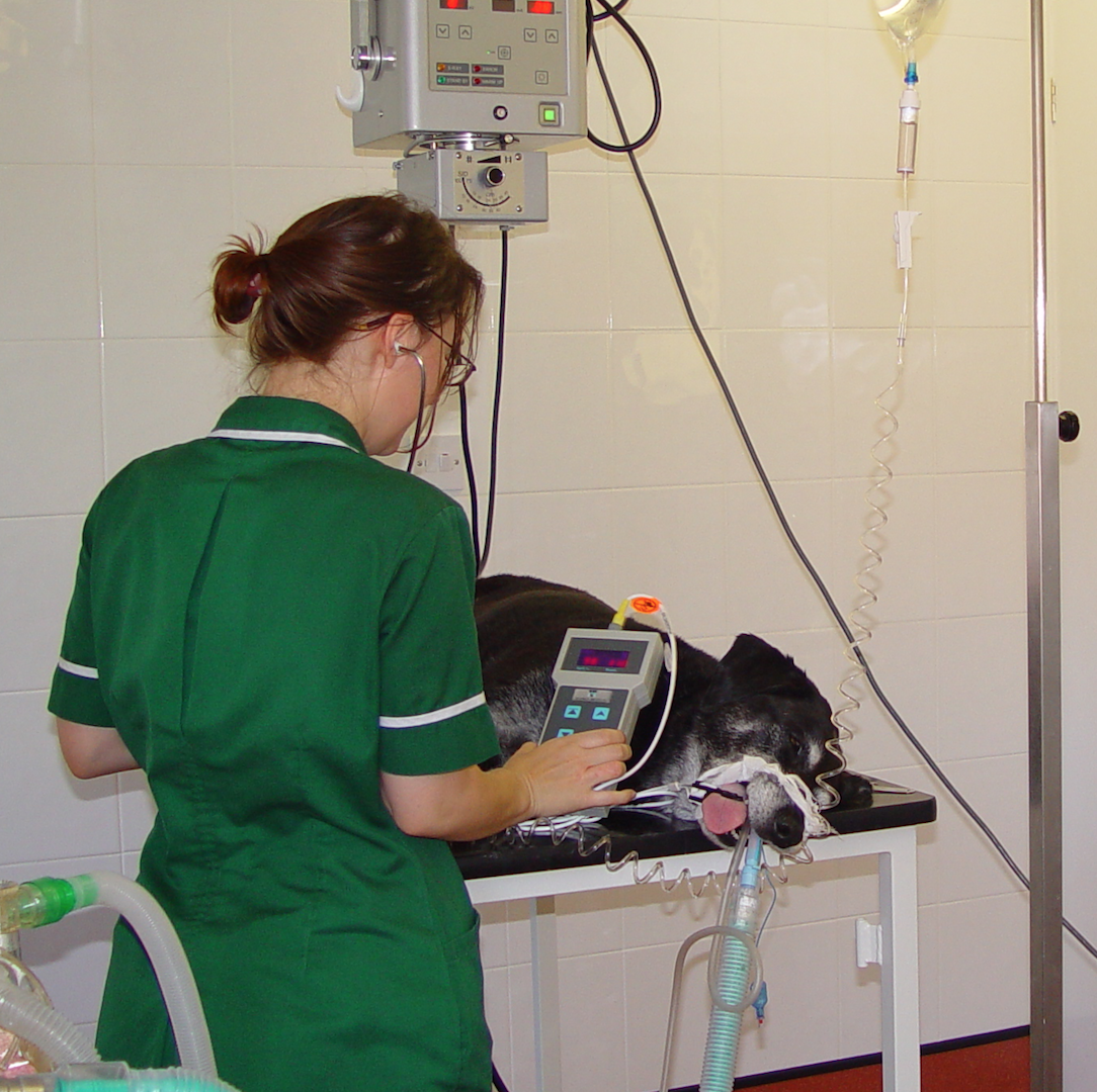
Before your pet has any anaesthetic your veterinary surgeon may discuss with you pre-anaesthetic blood tests, amongst many important considerations. All of the information provided to you is vital to ensure you are made aware of all of the risks and benefits to your pet associated with anaesthesia and surgery.
What is the aim of pre-anaesthetic blood tests?
The aim of performing pre-anaesthetic blood tests is to aid in detecting something that a full examination performed by your veterinary surgeon may not find. It is also a way of checking certain aspects of your pet’s health and may help in the future when looking at changes over time if your pet becomes ill.
What happens to my pet before they have an anaesthetic?
- Your veterinary surgeon will fully examine your pet as they would do in the consultation room
- Particular focus will be made to assessing the heart and lungs, which your veterinary surgeon will listen to using a stethoscope
- Any abnormalities detected during the general examination may lead your veterinary surgeon to advise additional tests, which may or may not include blood tests, xrays and ultrasound
- In the majority of cases where your pet is healthy and requires anaesthesia for a non-urgent procedure, nothing will be found during the clinical examination
- Pre-anaesthetic blood testing may be performed on the morning your pet is admitted, or prior to this if it is a non-urgent procedure
What does pre-anaesthetic blood testing look for?
- Illness or certain disease not detectable on routine examination of your pet. Organs such as the kidney and liver are routinely looked at using blood tests, as some changes will not always be detectable on general examination of your pet
- Presence of low red blood cell count (anaemia)
- Presence of cells suggesting infection or inflammatory disease (high white blood cell count)
- Evidence of dehydration (high red blood cell count and high total protein levels)
What information may pre-anaesthetic blood testing NOT give my veterinary surgeon?
- The cause of the body organ disease or dysfunction may not always be identified and further testing may be required if an abnormal result is detected
- Not all organs can be routinely tested for. Certain organ function tests require blood sample to be sent to an external laboratory and therefore need to be taken prior to any planned anaesthetic
What questions should I discuss with my veterinary surgeon regarding pre-anaesthetic blood testing?
- In which pets they recommend blood testing
- How the results of the blood testing will either change or impact on the management of your pet’s anaesthetic
- What will happen if the results are abnormal? For example would they still recommend an anaesthetic or would they recommend further tests prior to anaesthesia
When should my pet have their blood test?
Although blood testing may be performed by your veterinary surgeon, in the majority of cases this does not provide any time to correct or investigate any problems for a planned procedure. In an emergency or urgent situation then blood testing will, of course be performed in this manner.
Blood testing prior to your pet being admitted for their anaesthetic will allow for any further testing required to be performed and prevent any delay on the day itself. In most cases, blood testing is best performed with your pet starved to give the most accurate results.
How will my pet have their blood test performed?
Blood will be collected from a blood vessel, most commonly the jugular vein in the neck, with your pet held by a veterinary nurse. The sampling is performed in exactly the same way as a blood sample would be taken from yourself.
What evidence do we have when considering pre-anaesthetic blood tests?
Several studies have been performed in dogs to look at the benefits of pre-anaesthetic blood testing. These studies have helped to determine when the results may lead to a change in the management of your pet’s anaesthetic and therefore offer a benefit to your pet’s health. Importantly, even with these published studies the decision as to whether to perform any blood testing should be one you consider carefully with your veterinary surgeon.
The studies performed to date have shown that routine pre-anaesthetic blood testing does not impact on anaesthetic management in a healthy pet, undergoing a planned, routine procedure. It is important to note that when your pet is unwell or requires a particularly difficult or invasive procedure, blood testing prior to an anaesthetic will most likely benefit your pet’s treatment and allow your veterinary surgeon to plan effectively and safely. Only in dogs over the age of 8 years did blood testing help to detect disease that was not evident on clinical examination. There will always be cases though, where blood testing may detect something not found on full examination of your pet.
Recommendations on blood tests before anaesthesia
- All dogs over 8 years old are recommended to have blood testing prior to an anaesthetic. Although there are no similar studies in cats, blood testing should be considered when they over 8-10 years old
- If your pet is unwell and presents as an emergency to your veterinary surgeon, blood testing is recommended
- If your veterinary surgeon detects an abnormality on their examination of your pet, blood testing is recommended
- If you have any concerns regarding your pet’s health then you should discuss the benefits of pre-anaesthetic blood testing with your veterinary surgeon
Nasal discharge and sneezing in cats
by admin on January 4th, 2016
Category: News, Tags:

Cats with nasal discharge, “snuffly” breathing, snoring noises when asleep or awake, sneezing and sometimes gagging, are suffering from disease affecting the nose and/or throat. Signs that have been ongoing for more than 3 weeks are termed ‘chronic’, but many cats have problems for weeks to months, often with a variable response to treatment, before full investigations are carried out.
What causes nasal discharge and sneezing in cats?
There are many causes of nasal disease. The feline ‘flu viruses (herpesvirus and calicivirus), will commonly cause sneezing and discharge from the eyes and/or nose. There may also be a high temperature and loss of appetite. However the signs from these infections are usually short lived.
For recurrent or long-standing nasal discharge, antibiotics are often prescribed initially. However it is unusual for a bacterial infection to be the primary problem. There may be improvement with antibiotics as they will reduce secondary infection, but the signs are likely to recur when antibiotics are stopped. It is still important to address secondary infection though as, if it is left untreated, deep-seated infection in the bone (‘osteomyelitis’) may develop if the patient is unlucky.
Some primary causes of nasal discharge and sneezing in cats:
- Rhinitis (benign inflammation)
- Foreign body (often a grass blade)
- Fungal infection (uncommon)
- Benign polyp (an abnormal growth of tissue projecting from a mucous membrane)
- Tumour
- Severe dental disease
- Scar tissue narrowing the back of the nose
How will my vet investigate nasal discharge in my cat?
Your vet will want to know what type of discharge is present, as clear or yellow/green discharge is usual but if there is fresh (red) or old (brown) blood, this may suggest that a fungal infection or tumour is more likely.
Your vet may also want to know if the discharge is from one side of the nose or both sides, as tumours and foreign bodies typically affect one side only, whereas rhinitis usually affects both sides. It is important to tell your vet if your cat has stopped eating as this may be a sign of pain or an indication of a tumour.
Examination will usually include your vet assessing airflow through your cat’s nostrils by holding up a thin strand of cotton wool and looking for movement, or seeing if condensation forms on a shiny surface held close to your cat’s nose.
Your vet’s initial investigations are likely to include examination of your pet’s teeth, nose and throat under anaesthetic. A ‘polyp’ may be visible this way, and grass blades can sometimes be grabbed and removed quite easily if part of them is visible just inside the nostril or at the throat.
X-rays can be useful to look for tumours or destruction of the fine bone structure in the nose. Where available, computed tomography (CT) scanning gives even better detail than an X-ray. Endoscopy is very useful as this allows direct visualisation of your cat’s throat and nasal passages. Short or well embedded foreign bodies are visible with this technique and can be removed. Nasal tissue biopsies can also be collected with the aid of endoscopy, and biopsies are helpful to confirm rhinitis and essential to diagnose a tumour.
What treatment options are there?
Non-specific treatments such as steam inhalation and applying saline drops into the nostrils can help to loosen and clear discharge from the nasal passages. Other treatments will depend on the specific underlying cause:
- Rhinitis cannot be cured, but can usually be managed with a combination of anti-inflammatories and intermittent antibiotics. In many cases anti-inflammatory treatments may be best administered topically, via nasal drops or an inhaler as is used for asthma
- Fungal infection can be cured but treatment may take many months to be effective, and sometimes surgery is required to instil anti-fungal solution and cream into the sinuses
- Polyps can be removed with gentle pulling, but recurrence is possible. Surgery to address the primary site of growth in the middle ear is likely to result in permanent resolution
- Some tumours may respond to treatment, but sadly others are untreatable
Some causes of chronic nasal disease in cats can be cured, others can only be managed. However with appropriate investigations and treatment, a good quality of life should be achievable in the majority of patients.
Osteoarthritis in Dogs
by admin on December 2nd, 2015
Category: News, Tags:

What is osteoarthritis?
Arthritis means ‘inflammation of a joint’. Inflammation is a basic way the body reacts to infection, irritation or trauma and we generally see redness, swelling, warmth and pain. So ‘arthritis’ is actually not a very specific term and there are a number of quite different problems that will cause inflammation of a joint. However, the most common form of arthritis in dogs is called osteoarthritis (“OA”). This is sometimes called rheumatism, osteoarthrosis or degenerative joint disease (DJD).
What happens in osteoarthritis?
In osteoarthritis there is gradual deterioration and loss of the smooth cartilage surface of the joints. The soft tissues around the joint can become inflamed and the joint gets thicker and stiffer. This form of arthritis in dogs often develops slowly and can be well advanced before the joint becomes sore.
What causes osteoarthritis?
There are then a number of potential TRIGGER factors for osteoarthritis but once triggered, osteoarthritis appears to be a SELF-PERPETUATING PROCESS that generally progresses at a variable rate. So once a patient has it, they have it! Osteoarthritis in dogs is usually triggered by another joint problem such a Hip Dysplasia, Elbow Dysplasia and Osteochodrosis or by trauma to the joint.
Osteoarthritis is the form of arthritis typically associated with getting older and with wear and tear. However, if a growing dog has a developmental problem such as Elbow Dysplasia or Hip Dysplasia then osteoarthritis will develop in a joint at a very young age. What’s also interesting is that many older people and dogs will show little or no sign of arthritis, even if they have been very active and their joints have worked hard. So it is not just an inevitable consequence of aging; there are other factors at work.
What are the signs?
Stiffness after rest is a characteristic feature of osteoarthritis and these usually starts quite subtly. In some patients, quite marked lameness can come on suddenly, usually after particularly vigorous or unfamiliar types of exercise. These signs can be caused by other conditions that may require completely different treatment and so the diagnosis can’t be made on symptoms and signs alone. You may find your dog spend more time in their basket and maybe starts to struggle getting in and out of the car.
How is it diagnosed?
Osteoarthritis is usually diagnosed from the typical symptoms and signs exhibited by the patient. X-rays will be taken to confirm the characteristic bony changes around the affected joint(s). Your dog will be sedated or anaesthetised for xrays and this is a very convenient time to take a sample of joint (synovial) fluid for laboratory analysis. This can help to rule out other forms of arthritis such as rheumatoid arthritis or a bacterial infection in the joint. In some patients a camera may be used for direct visual examination of the inside of the affected joint. This is called arthroscopy and it is a type of key-hole surgery.
How is it treated?
There are three steps in treating osteoarthritis in dogs.
Firstly we should look at any lifestyle modifications that could help. By focusing on some relatively simple steps, you can have a dramatic effect on your pet’s mobility and quality of life.
In the early stages it is likely that the patient will receive little specific medical treatment and in the early stages we should focus on preventing the condition worsening and avoiding things that may aggravate the joint and make it more painful. In general, exercising on flat even ground is going to be kinder to all the joints. Exercise within the capability of your pet. This will be trial and error as osteoarthritis is a very variable condition and what is ok for one patient may not be suitable for another. Don’t exercise your pet to a level that aggravates signs and symptoms. It is essential that your pet gets down to an appropriate body weight. I can’t emphasise this enough. In people, it is not proven that being overweight will cause osteoarthritis – it may be one factor that could contribute in some patients. However, being overweight will definitely make your symptoms worse and increase your dependency on medications to control joint discomfort.
Secondly, a course of anti-inflammatories may be required to ‘settle’ the joint. Some patients require long-term medication with these drugs. Using a nutritional supplement such as glucosamine and chondroitin sulphate may be beneficial and some patients will be helped by a course of Cartrophen injections.
Thirdly, surgery may be required in some patients. The main role of surgery is in the management of established osteoarthritis to replace a worn-out, chronically painful ‘end stage’ joint. None of us wants are pet to undergo surgery anymore than we want it ourselves but appropriate surgery at the right time can be life changing for dogs with severe, painful osteoarthritis. The main types of surgery performed are joint replacement, arthrodesis and excisional arthroplasty.
How Can We Tell if Dogs and Cats are In Pain?
by admin on November 2nd, 2015
Category: News, Tags:

How can we tell if dogs and cats are in pain? It’s a very good question! In this article, we explain the importance and some of the challenges to providing adequate pain relief for dogs and cats.
Do animals feel pain like us?
Yes they do. However, for many years this was questioned. The International Association for the Study of Pain (ISAP) defines pain as both a sensory and an emotional experience. In other words, as well as feeling the ‘ouch’ part of pain, there is also an emotional aspect to it. That is why we often feel unhappy if we experience pain. Your back is sore, it hurts to do all the things you do normally and this affects your mood and motivation. This is true for our pets as well and most pet owners have a fair idea when their animal is not right and seems a bit down. Even so, quantifying how a pet’s pain affects them is difficult because they are limited in the ways they can communicate to us. The situation is similar in babies, the elderly and critically ill people – the ability to communicate pain is affected.
The IASP definition of pain goes on to say that “the inability to communicate verbally does not mean that an individual isn’t experiencing pain and that they don’t need appropriate pain-relief”. In fact, with our patients we nearly always err on the side of caution and administer a pain killer if we are unsure. The response of the animal will often confirm we made the right decision!
Pain is an individual experience. We often hear people say that someone has a high (or low) pain threshold. No two people or animals react in exactly the same way to a given painful stimulus. This is because the pain processing systems in our body are not uniform but adapt based on previous experiences. If we experience a lot of pain over time we seem to get sensitised to it. This is one reason why pain control after surgery for example is so important. It can speed up you feeling better. There is good evidence that babies who undergo painful procedures are more sensitive to pain in adulthood and the same can be said of animals.
Have you ever been asked to score your own pain?
There has been much work in recent years into pain scoring (or simply ‘putting a number on it’) in dogs and cats. If you undergo an operation in hospital your doctor will ask you whether you experience pain. Sometimes you will be asked to say how bad it is on a scale of 1 to 10. This ‘scoring’ system is extremely valuable for doctors not just to get a better idea of just how bad it seems to you, but also for assessing your response to pain killers or other treatments – they will look to see if your pain score is decreasing.
Pain assessment is not as easy in dogs and cats
For dogs and cats we cannot simply ask them how painful they are. Well, we can but the responses may not be so reliable! Therefore we need to develop reliable systems to gauge their pain from their behaviour.
Such scoring systems have been used widely for dogs and are currently being developed for cats – cat behaviour is a bit more difficult to work out. We use pain scoring after surgery to guide our pain management in the post-operative period. In addition to this, experience with the procedures we commonly perform also gives us the knowledge to know what to expect.
Isn’t a little bit of pain a good thing?
Our ability to treat pain as vets is increasing steadily as new drugs become available, combined with systems designed to evaluate pain in dogs and cats. Educating pet owners also plays a big part. Whilst some owners may consider it normal for a dog to limp – because that’s what their old dog did – this should no longer necessarily be accepted as ‘just getting old’.
There are various pain management options for both dogs and cats long term which allow us as vets and owners to improve the quality of life of our pets. In the past you may have heard people say that a little bit of pain is a good thing. For example, it stops your pet from moving around too much after surgery. This is certainly not true and we have good evidence that too much pain can be detrimental to recovery from surgery. If you consider your pet to be in pain after surgery, then you should ask your vet to prescribe pain killers or at the very least take them in for a check up.
What can we do at home?
Your role in working out the degree of pain your pet is experiencing is just as important as that of your vet. Another way you can help is completing pain scoring charts when you visit your vet. Now, we don’t use these all the time with owners but they can be very helpful if your pet is having regular visits to assess response to treatment for conditions such as arthritis.
These pain scoring systems which we may ask owners to complete at each visit to assess the overall picture and do not solely base assessment of pain on our examination of the pet in the clinic. You know your pet better than anyone and often in a consulting room some dogs and cats will just freeze and not show any pain. We see a different picture to the one you see at home..
Dog Aggression to Owners
by admin on October 2nd, 2015
Category: News, Tags:

If a dog is showing threat towards or biting family members it is perhaps one of the hardest problem behaviours to live with. Not only does it place the family at constant risk, but it can also be very hard to understand why a dog that is loved and well cared for behaves this way. So why do dogs sometimes bite the hand that feeds them?
As discussed in the last month’s article ‘Dog Aggression’ there are many reasons why dogs sometimes use threat or aggression, and all of these apply equally in cases of dog aggression to owners. However there are some particularly common scenarios in which dogs may use aggression towards family members, many of which can be prevented.
Puppy development
Puppies, like children, pass through numerous periods of development before they reach maturity. One of the most rapid of these occurs between 3 and about 12-14 weeks of age. This is when puppies start to explore and learn about the world around them. They are particularly sensitive to forming bonds with other species, such as humans or other family pets, learning what is safe and developing the skills to cope with things that bother them during this period. It is therefore important this is managed carefully.
However behavioural development doesn’t stop there. As the puppy continues to grow he will go through a series of further behavioural developmental stages. For example some puppies experience a period of increased sensitivity to potentially scary experiences, typically somewhere between 9 -18 months. If this isn’t handled in the right way it can lead to longer term fearfulness, which can in some cases lead to aggressive behaviour due to fear or panic.
There are also changes linked to fluctuating hormone levels, just as in teenage humans. Testosterone is linked to confidence, the drive to breed and the dog’s willingness to take risks. It can therefore increase the likelihood a dog will challenge for something he wants, the speed with which he goes from warning to biting and how much force he uses. Testosterone levels start to rise as young male puppies approach puberty and can fluctuate until they settle down as the puppy matures. Owners may therefore find their puppy is particularly confident or pushy at times and occasionally even tries things like growling to see if it gets him his own way during this period. If this is handled calmly it usually passes. However if the puppy either learns it works or is punished for it then this behaviour may get worse over time.
Bitches also start to be affected by their hormones with the onset of their seasons. They can be more irritable and competitive during the early part of their season and this may lead to uncharacteristic snappy behaviour. This also usually settles as the season passes, as long as it isn’t aggravated, but may recur again the next time.
Behavioural development continues until the dog reaches maturity at between 18-36 months, depending on the breed and the individual. This process involves ongoing changes in the brain with existing nerve cells constantly being replaced by new ones. Once a dog reaches maturity they often become calmer and more restful. However, in some cases maturity may also lead to a change in the way the dogs react to things that bother them. For example a puppy that uses appeasing to ask someone not to touch them in a way he finds worrying, may escalate to using threat when this doesn’t work once they are mature.
Defensive aggression
Another common trigger for dog aggression to owners is fear. This is most commonly caused by excessive use of punishment. If a dog learns that a certain way of behaving leads to something he or she doesn’t like, such as being ignored or the owner leaving the room, this can deter unwanted behaviour and teach better manners. However harsher punishments (see Box 1) may make the dog fearful or cause pain and so trigger defensive behaviour. This is particularly likely to happen if these types of punishment are given at the wrong time or aren’t given consistently. The dog won’t be able to work out the reason for them and so starts to expect them to happen at any time. The dog then resorts to using defensive aggression to try and prevent them. This may make the dog seem unpredictable. However, the dog is in fact reacting to what they see as unpredictable behaviour by the human and so are only doing what they feel they have to do to protect themselves from anticipated punishment.
Punishment methods that may trigger defensive aggression:
- Smacking
- Hitting the dog with a rolled up newspaper
- Alpha rolling (forcing the dog onto his or her side)
- Shouting or staring at the dog
- Grabbing the dog by the scruff
- Jabbing the dog with a foot, fingers or clawed hand
- Collars that tighten around the dog’s neck e.g. check chains, slip leads and prong collars
- Use of a lead to correct or force the dog to do something
- Electric ‘shock’ collars
- Rattling cans of stones, spraying with water or using canisters that emit a hiss of air in dogs that are worried by them
Being groomed
Dog aggression to owners can also occasionally arise when a dog is groomed or handled in a way he or she doesn’t feel comfortable with. This can be because the dog is in pain, or has learnt to associate being groomed or handled with pain. It can also be due to fear if the dog has been punished for wriggling in the past. Some of the ways humans handle dogs can also seem threatening to them, even if they are not intended to be. For example dogs threaten each other by putting their head over the other’s neck and so a worried dog may see a human touching the back of their neck as a threat. Some dogs also simply don’t like being touched on certain parts of the body such as their feet or under the tail. This can result in the dog using threat to ask the person to stop.
Children
Dogs and children can be of great benefit to each other and dogs generally instinctively know to be gentle with children and babies. However, every dog has its limits and children, especially younger ones, can sometimes unintentionally do things to dogs that hurt or scare them. Lots of noisy children can also encourage dogs to become over excited which can then sometimes spill into overly boisterous behaviour or – rarely – aggression, just as an over excited child may have a tantrum. Providing suitable escapes for dogs, making sure they get enough rest, teaching children how to behave around them and supervising young children around dogs can all go a long way to preventing this type of problem behaviour. You can find more information about how to keep dogs and children safe together in the article ‘Children and Dogs’.
What should I do if my dog shows threat towards me or bites me?
All of these types of aggression can be prevented, or corrected with the help of an accredited behaviourist. If your dog is showing aggression to you the first port of call is your veterinary surgeon, so he or she can be checked for any signs of illness or pain. If your dog is given a clean bill of health your vet can then refer you to an accredited behaviourist who can assess the reason for the problem behaviour and advise you how to address it without making it worse.
