News
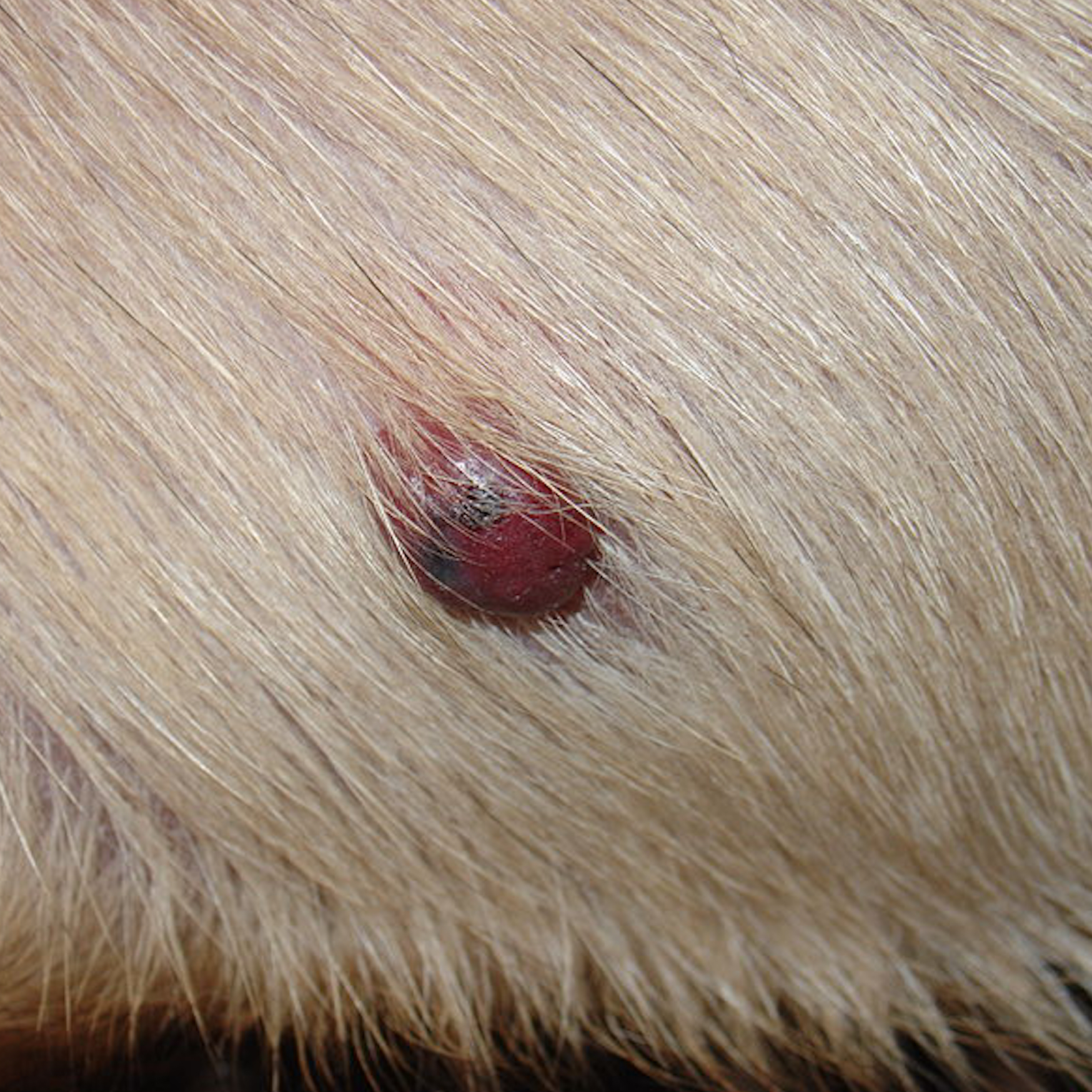
Cancer in cats and dogs
by admin on February 1st, 2019
Category: News, Tags:
Assessment of the patient
Cancer is common in human and veterinary medicine. Approximately one quarter to one third of all our patients will suffer from cancer at some point in their lives, which is similar to the incidence of cancer in human medicine. So, we encounter a large number of patients with cancer during our clinical work, and we also find that many owners themselves will have some first-hand knowledge of cancer, which may affect the decisions they make about the disease in their pet.
The investigation and management of animals suspected of having cancer or known to have cancer is relatively straightforward and generally proceeds in a similar manner for all patients, with a few basic rules followed in a stepwise manner. When investigating a particular tumour, we never forget that the tumour is attached to a patient, and that we need to carefully consider the care and needs of the pet as well as those of their owner. Our approach is one of keeping owners fully informed at each step and making plans for investigation and management of the pet only after careful discussion with the owner.
Outline:
The following information is divided into the following sections:
- Investigation and care of the patient
- Investigation of the tumour
- Identification of the most appropriate therapy
The information is arranged in this way, because this is the way that our patients present to us and because we think of the patient first, his or her disease second and the proposed therapy third. In particular, we always make every effort to ensure that we have a correct diagnosis before we begin therapy and that the patient will be able to tolerate the treatment prior to us starting it.
1. Investigation of the patient
General assessment of the patient
When presented with an animal suspected of having or known to have cancer, our initial approach is to assess the patient and then assess the tumour. Each patient is different, as is each tumour, and the investigative approach is therefore tailored to the individual, although the same basic principles apply.
When presented with a patient with cancer, we want to get an accurate assessment of the long-term outlook or prognosis. The prognosis for animals with cancer depends on a number of factors:
- The microscopic or histological type of the tumour, i.e. what cells are in the tumour?
- The grade of the tumour, i.e. how aggressive do these cells look under the microscope?
- The clinical stage of the tumour, i.e. is there any evidence that the tumour is affecting more than one site in the body?
- The presence of tumour-related complications (so-called ‘paraneoplastic syndromes’)
- The presence of other diseases
Our approach is to:
- Assess the general health of the patient
- Identify any other diseases present
- Confirm the presence of a tumour
- Gain as much information as possible about the tumour
- Identify how the tumour is affecting the patient currently
The basic principles in assessing the patient with cancer comprise:
- Tumour measurement
- Staging the tumour
- Assessment of the patient’s general health and fitness
- Diagnostic imaging (e.g. X-rays, ultrasound, CT scan, MRI scan)
- Blood samples to look at the various blood cells and other substances in the blood (haematology and biochemistry)
- Examination of samples from the tumour or other sites, either samples of cells (cytology) or tissue (histology)
Two important factors are the grade and stage of the tumour, both of which must be assessed. Grade is assessed under the microscope and compares the tumour cells to the normal cells of the same type.
- Grade predicts the biological behaviour of the tumour, e.g. benign or malignant and how different from normal the cells look
- Grade answers the question ‘How might the tumour affect the patient in the future?’
Stage is assessed clinically and with diagnostic imaging and sometimes samples of cells or tissues.
- Stage tells us where the tumour is within the patient, e.g. is it restricted to the site where it started growing, or has it spread to other tissues, e.g. lymph glands (lymph nodes) and other organs
- Stage answers the question ‘How is the tumour affecting the patient right now?’
Tumour staging
One of the most important ways to assess the patient with cancer, is the ‘TNM’ system. This involves:
- Examination of the primary Tumour (with a biopsy – see later)
- Assessment of the regional lymph Nodes (the ‘glands’ such as we have in our necks)
- Assessment of other sites for Metastasis (spread elsewhere)
Staging is generally performed by a combination of physical examination, diagnostic imaging and taking biopsy samples of the tumour to look at under the microscope (‘cytology’ or ‘histology’). In some cases, the appearance on diagnostic imaging may be sufficient to make a diagnosis without biopsy, e.g. nodules in the lungs seen on an X-ray of the chest suggests spread of cancer cells to the lungs.
This clinical staging represents the ‘gold standard’. However, this approach is not necessary or appropriate in all cases. For instance, for a pet with a benign tumour confirmed with a biopsy taken by sucking cells out of the tumour through a hypodermic needle (needle biopsy), the prognosis (outlook) is excellent and the likelihood of tumour spread is very low, so staging may be taken no further after gaining some information about the primary tumour.
Care of animals with cancer
We all may have our own view of how the tumour is affecting the pet and owners may well have other concerns about their pets apart from the fact that they have a tumour. The supportive care of the patient is the second part of compassionate patient care, after ‘diagnosis’ and before ‘therapy’. This is important, since we wish to avoid the tendency to leap from diagnosis to treatment without considering what supportive care is needed.
Our approach can be summarised by three important rules, which we all consider:
- We should ensure that our patients do not experience pain or discomfort associated with the tumour or its treatment
- We should ensure that our patients are able to eat a normal amount of food to meet their nutritional requirements, and if they cannot, we should think about how to help them to do so
- We should ensure that our patients do not suffer side effects of the tumour or therapy, such as vomiting or feeling sick when undergoing treatment, if at all possible
Management of chronic cancer pain
When asked what our concerns about cancer would be, most of us would say that pain associated with cancer would be uppermost in our minds. As a rule, we understand how to manage acute pain after an operation (see later), but the management of chronic cancer pain in a patient where a total cure is not possible is sometimes more difficult to achieve.
We have detailed methods of assessment of pain and discomfort and understand the importance of treating and managing any pain or discomfort associated with cancer. It is important to realise that management of pain does not just involve drugs traditionally thought of as ‘painkillers’ but may also include other drugs that have an effect on the way nerves and the brain deal with pain (e.g. gabapentin and amitryptiline) or drugs and other therapies that have an effect on the tumour itself (e.g. bisphosphonates or radiotherapy in cases of the bone tumour, osteosarcoma).
We should also bear in mind that in some tumours the most successful method of pain relief may be surgical removal of the tumour itself, whether that is a procedure designed to cure the patient or a treatment designed to remove or reduce the clinical signs associated with the presence of the tumour without totally curing it.
2. Investigation of the tumour
Tumour biology
As a general rule, if we do not understand the basic biology of a disease i.e. the behaviour of the cancer cells in the patient’s body, we cannot provide appropriate treatment. One important lesson that has emerged from both human and veterinary oncology over the past few years is that, when it comes to cancer, the biology of the disease will dictate what happens in the long-term.
A common question asked when a diagnosis of cancer is made in our pet is ‘What caused the cancer?’ While we know something about some causes of cancer (and what is happening in the cancer cells and their genes), for the most part, we do not know the cause.
The total number of cells in a person or animal is a balance between the number of cells growing and those dying. For a long time it has been considered that cancer is a disease caused by uncontrolled cell growth. However, of more importance is the fact that cancer cells refuse to die when programmed to do so (this programmed, and perfectly normal, cell death is called ‘apoptosis’). Cancer therapies that encourage cell death are likely to be just as important as, if not more important, than therapies that prevent cell growth.
Other examples of cutting edge treatments in humans that have been developed as a result of understanding tumour cell biology include those designed to stop the early phase of tumour spread (metastasis) by preventing tumour cells moving through the tissue, and those causing a reduction in tumour growth by preventing the tumour causing the growth of new blood vessels (angiogenesis) which the tumour needs in order to grow.
Tumour pathology
Pathologists play a critical role in the investigation of the patient with cancer by examining tissue from the primary tumour (the original site of the growth) and, in some cases, the lymph nodes and other potential sites for spread (metastasis).
Assessment of cells obtained from a mass with a hypodermic needle (cytology) is often the first approach in the diagnosis of an animal suspected of having cancer, but in most animals, examination of a tissue sample (histology) is necessary at some point during the assessment of the patient.
Histological assessment of the tumour may also provide more specific information regarding the outlook or prognosis:
- Examination of the tumour edges after the growth has been surgically removed may tell us whether the tumour is likely to grow back in the same place (i.e. if there are tumour cells at the edge of the specimen, it is likely that tumour cells have been left in the patient)
- Examination of a large specimen for evidence of tumour cells spreading into the blood vessels or lymph drainage vessels may suggest whether the tumour is likely to spread
Other effects of the tumour (Paraneoplastic syndromes)
Paraneoplastic syndromes are additional effects that cancer might have in parts of the body away from the primary (or original) tumour. These abnormalities may have an effect on the patient’s health and may affect their ability to tolerate sedation or anaesthesia and treatment. The paraneoplastic syndrome may be the main reason for the animal being taken to the veterinary surgeon in the first place (the owner may not even realise that there is a tumour somewhere) and these syndromes also may provide a useful guide to the success of therapy, i.e. if the paraneoplastic syndrome gets better, this suggests that the tumour tissue has been removed or destroyed. In rare cases, the paraneoplastic syndrome may be very obvious but the location of the underlying primary tumour is more difficult to find, even for the veterinary surgeon. In such cases, additional imaging procedures can prove very helpful.
Examples of these syndromes are alterations in the number of red blood cells or white blood cells in the circulation and abnormalities in the blood level of glucose or certain salts or ‘electrolytes’ (e.g. calcium).
3. Treatment of the patient with cancer
Achieving a diagnosis
As a general rule, a biopsy (taking cells or a larger piece of the tumour for analysis) is needed before beginning any therapy, unless we have a good reason not to take a biopsy. A biopsy may be omitted if knowing more about the tumour would not change the treatment plan or the willingness to treat the patient. However, care is taken to ensure that these reasons apply in any individual patient. A biopsy should always be tailored to the individual tumour and we will always consider whether a biopsy of the regional lymph node (gland) or other sites of potential metastasis (spread) is needed at the same time.
Selecting the best cancer treatment for the patient
Once a diagnosis has been achieved and we have information on clinical staging, the effect of the tumour on the patient and any concurrent diseases, and once we have identified whether the patient needs additional supportive care, we can start to consider different treatment options.
The main treatment options for the management of cancer are:
- Surgery: The physical removal of the tumour
- Radiotherapy: The use of a strong X-ray beam to destroy the cancer cells where they sit
- Chemotherapy: The use of anti-cancer drugs to kill cancer cells wherever they are
Each of these has specific advantages and disadvantages and may be used singly or in combination. All of the factors listed above, particularly the tumour type, grade, stage and location and the presence of other health problems will help to decide what the most appropriate therapy is. Therapy may be designed to cure the patient or to remove the signs of illness for as long as possible, if a complete cure is not possible.
In addition, other ways of treating various types of cancer are becoming more common in human oncology, and these benefits will become available for our patients in due course.
These include:
- Immunotherapy, e.g. vaccination with melanoma-specific antigens in patients with melanoma (a tumour of the pigment cells in the body)
- Targeted therapy with small molecule inhibitors, e.g. tyrosine kinase inhibitors (TKIs) which may be used in some patients with mast cell tumours
Monitoring the response to therapy
Once the cancer has been treated, the patient will still be monitored in various ways, depending on the nature of the tumour and the aim of the therapy. This may involve a number of the tests (e.g. blood samples, X-rays) used to make the diagnosis in the first place and a plan for monitoring will be tailored to the needs of the patient. This may include:
- Monitoring for the return (local recurrence) of the tumour at the original site
- Examining for the presence of tumour spread elsewhere
- Monitoring for the development of new primary tumours at other sites
If you have any questions about your pet’s condition, or his or her treatment, please do not hesitate to contact us.